Lupine Publishers | Advancements in Cardiology Research & Reports
In this review, we hope to give a perspective of the new realities of
cardiac mechanical circulatory assist devices. New
iterations of devices are providing greater durability and freedom of
complications. Work is near to provide internal batteries and
transcutaneous energy transfer systems for completely implantable
systems, avoiding the need for an externalized drive line.
Keywords: Heart Failure; Transplantation; Mechanical Circulatory Support
Twine and Twine or Lose the Plug- Dislodged Left
Atrial Appendage Closure Device
In patients with severe heart failure, cardiac transplantation has
been shown to provide considerable benefit. Since 1967, in excess
of 88,000 total heart transplants have been performed and 1-year
survival is 81%, the annual mortality is 4% per year thereafter. The
supply of donor hearts is incredibly limited and much research has
focused on mechanical means of improving myocardial function,
and several such left ventricular assist devices (LVADs) have been
developed through the National Institutes of Health artificialheart
program. Several devices have been previously approved
by the Food and Drug Administration as bridging therapy to
transplantation, though none have been studied as long-term
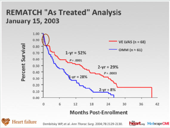
alternatives to transplantation. The Randomized Evaluation of
Mechanical Assistance for the Treatment of Congestive Heart Failure
(REMATCH) [1] trial explored whether a specific type of LVAD (a
previous generation pulsatile device), when used in the long-term,
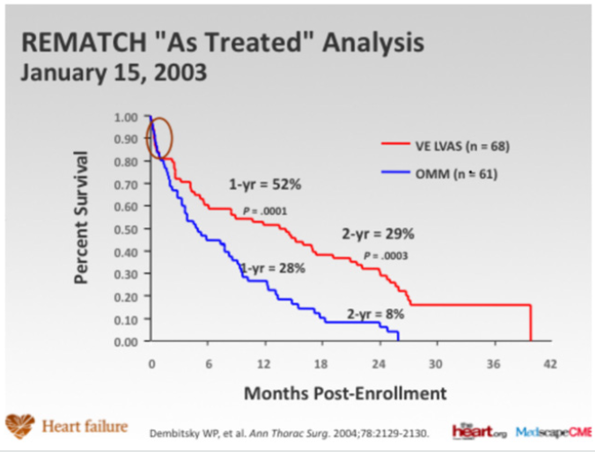
would reduce mortality (Figure 1). The survival following severe
heart failure was extremely poor in the optimally medically treated
group in this trial (defined as End-stage heart failure was defined as
New York Heart Association (NYHA) class IV symptoms for at least
90 days, left ventricular ejection fraction (LVEF) <25%, peak oxygen
consumption <12 mL/kg/min or continued need for intravenous
inotropes for symptomatic hypotension).
In the optimally medically treated control arm of the
Randomized Evaluation of Mechanical Assistance for the Treatment
of Congestive Heart Failure (REMATCH) trial [1] which evaluated
an externalized pulsation ventricular assist device, survival at one
year was 28% and 6% at two years, underlying the poor prognosis
of this clinical entity (Figure 1). Driving the need for mechanical
circulatory support (MCS) is the relative paucity of donors and the
unmet need for orthotopic heart transplantation in the general
population. There has also been an increase in the number of
patients who require mechanical circulatory support (MCS) as a
bridge to transplantation [2]. This has been driven, particularly
in the UK by limitation of the number of hearts for donation,
and also to buy time on the transplant waiting list. This is due to
an increase in the numbers of non-heart beating donors (DCDs),
whereby retrieval takes place in a circulation arrested donor,
and the increased survival of head injury patients and those with
intracranial bleeds who are treated by a decompressive craniotomy,
reducing the pool of donors who have raised intracranial pressure
and who have coned, resulting in brain stem death. The net result
is a retrieval rate for heart transplantation of around 19%. The
risk of having preformed antibodies directed against the donor
heart (sensitised patients) is increasingly likely and is particularly
challenging as it may increase the risk of rejection and allograft
vasculopathy. There has also been an increase in the number of
patients requiring MCS as a bridge to transplantation [3]. This
allows many severely ill adults and paediatric patients to survive
until a suitable donor heart is available. Patients with MCS are at
increased risk for rejection, infection, stroke, and bleeding. The
need for transfusions also increases the risk of pre-sensitization
[3-5]. Survival at 1 and 5 years is decreased in patients requiring
MCS prior to transplantation, but still higher than 80% and 70%,
respectively (ISHLT database) [2].
Figure 1: Randomized Evaluation of Mechanical Assistance for the Treatment of Congestive Heart Failure (REMATCH) trial
(Rose et al N Engl J Med 2001; 345:1435-1443).
Advances in Donor Allocation and Selection
Recipient criteria for heart transplantation include, severe
symptoms despite maximal medical management, the absence
of reversible or surgically amenable heart disease, and where
estimated 1-year survival is less than 50% [6]. An estimate of
functional capacity in ambulatory patients can be best quantified by
measurement of peak O
2 consumption (VO
2max). Patients with low
VO2max (<12 ml/min/kg) have high mortality even if treated with
beta blockers and transplantation should be considered for these
patients. In addition, heart failure prognosis scores to estimate
survival, such as the Heart Failure Severity Score may be used.
This calculates a survival probability on the basis of the presence
of ischaemic cardiomyopathy, resting heart rate, left ventricular
ejection fraction, mean blood pressure, interventricular conduction
delay, VO
2max and serum sodium concentration [7].
Figure 2: Competing outcomes for continuous flow LVADS (82% survival at 1 year, intention to treat).
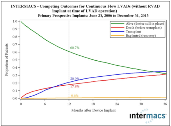
Transplantation eligibility is always considered with regard to
risk factors, especially, pulmonary hypertension (Figure 2). Right
heart catheterization must be performed in all potential candidates
for heart transplantation in order to quantify pulmonary vascular
resistance [7]. Right heart failure is a substantial cause of mortality.
Right ventricular failure is likely when post implant pulmonary
artery pressures exceed 50 mmHg. Patients with chronic heart
failure may develop pulmonary hypertension due to elevated left ventricular end diastolic pressure with elevated left atrial
and pulmonary venous pressures. This is a reactive form of
pulmonary hypertension and may fall when the cardiac output is
increased with inotropes or unloaded with nitrate infusions [7].
The transpulmonary gradient is calculated by subtracting the left
atrial filling pressure from the mean pulmonary artery pressure. A
fixed transpulmonary gradient in excess of 14 mmHg is associated
with greatly elevated risk, and thus this cut off is used in the UK [8].
In such patients a destination therapy strategy may be used with
continuous flow LVADS.
Mechanical Circulatory Assist Devices
In recent years, the use of MCS device in treating patients with
end-stage heart disease has increased significantly, as bridge to
transplantation and as destination therapy for transplant ineligible
candidates. This increase is based on the accumulated experience
with new second-generation continuous-flow devices which show
significant improvements in survival, functional capacity and quality
of life [9,10]. On the basis of the Heart Mate II Registry experience
(1300 patients), guidelines for the clinical management of patients
treated with continuous-flow devices have been published [11].
Risk scoring systems, such as the Seattle Heart Failure Model [12]
and the Cumulative Risk Score for 90-Day in-Hospital Mortality
[13] and the Destination Therapy Risk Score have been investigated
to stratify patients who might benefit from LVAD support [14].
Right ventricle failure is a leading cause of morbidity and
death after LVAD implant (incidence of about 35%), and can be
very difficult to predict [15,16]. Various means to assess right
ventricle function both pre- and postoperatively have been
assessed (10). Right ventricular failure risk scores have been
created that stratify the risk of right ventricular failure (RVFRS)
and death after LVAD implantation (Figure 3). One such RVFRS
found independent predictors of right ventricular failure to include
vasopressor requirement, aspartate aminotransferase >80 IU/L,
bilirubin >2.0mg/dL and creatinine >2.3mg/dL [15]. Another study
developed a score to predict RVAD need after LVAD placement,
which included factors of cardiac index, right ventricular stroke
work index, severe preoperative right ventricular dysfunction,
creatinine, previous cardiac surgery and systolic blood pressure
[16]. More recently the presence of severe TR and a tricuspid annulus
of >43mm and right ventricular sphericity have been proposed as
predictive of occult RV failure and need for biventricular support.
The Interagency Registry for Mechanically Assisted Circulatory
Support (INTERMACS) registry, which follows all long-term MCS
systems in the United States, has defined patient profiles that can
help identify risks associated with the timing of implant [17]. In
the future, the INTERMACS patient profile would be a useful tool
to improve management and outcomes of patients who need VAD
implant and unify criteria for future clinical trials and devices
(Figure 4). As more LVAD patients are listed for heart transplant,
a competition has occurred for organs between stable LVAD
supported registrants and less stable registrants listed UNOS status
1A or 1B (the highest categories and most at risk if not urgently
transplanted). A recent study found that stable LVAD patients had
significantly less 30-day risk of events compared to other status 1A
patients concluding that allowance of 30 days of elective status 1A
time should not be allocated to stable registrants with implanted
LVADs [18]. As VAD technology improves, further revisions to the
allocation system will need to be recommended.
Figure 3: Heartmate 3, the latest centrifugal blood pump in comparison to Heartmate II an axial flow pump. Superior event
free survival is seen with HM 3.
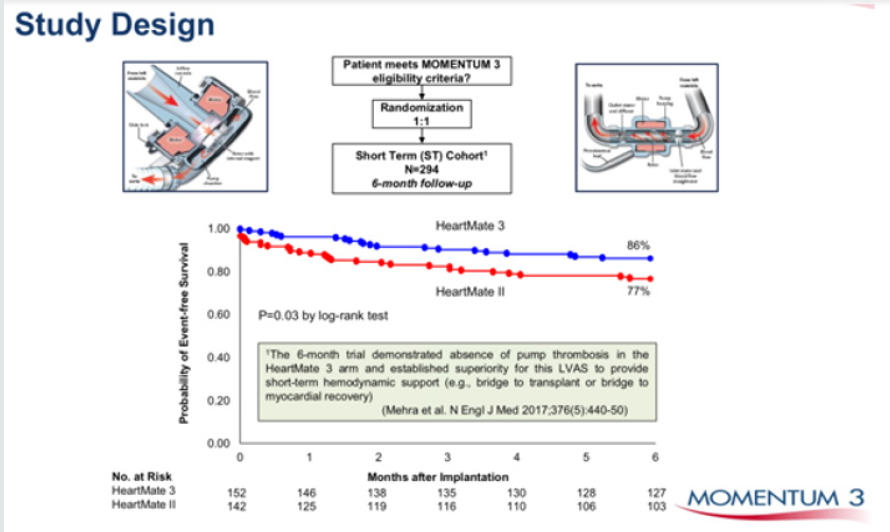
Figure 4: Heart-mate 3 vs Heartmate II comparison of event free survival.
INTERMACS Profile and Description and Timescale to MCS
a) “Crashing and burning”—critical cardiogenic shock.
Within hours
b) “Progressive decline”—inotrope dependence with
continuing deterioration. Within a few days
c) “Stable but inotrope dependent”—describes clinical
stability on mild-to-moderate doses of intravenous inotropes
(patients stable on temporary circulatory support without
inotropes are within this profile). Within a few weeks
d) “Recurrent advanced heart failure”—“recurrent” rather
than “refractory” decompensation. Within weeks to months
e) “Exertion intolerant”—describes patients who are
comfortable at rest but are exercise intolerant. Variable
f) “Exertion limited”—describes a patient who is able to do
some mild activity but fatigue results within a few minutes of any
meaningful physical exertion. Variable
g) “Advanced NYHA III”—describes patients who are
clinically stable with a reasonable level of comfortable activity,
despite history of previous decompensation that is not recent. Not
a candidate for MCS
INTERMACS = Interagency Registry for Mechanically Assisted
Circulatory Support; MCS = mechanical circulatory support;
NYHA = New York Heart Association.
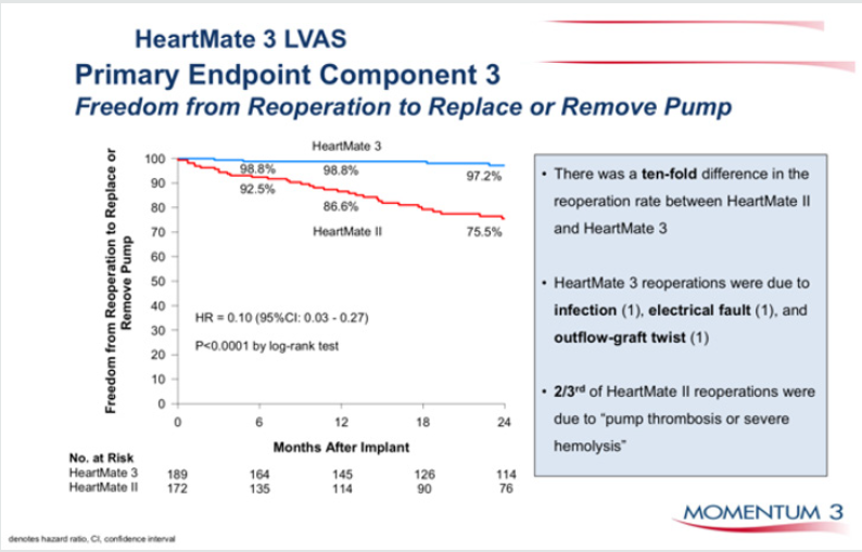
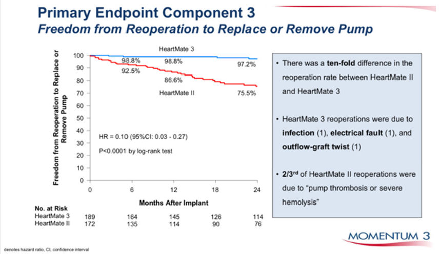
Figure 5: Durability of HM 3 vs HM II, freedom from pump replacement due to pump thrombosis or haemolysis.
Temporary MCS are available that can be implanted quickly and
simply to normalise cardiac output in patients with severe acutely
decompensated heart failure. The CentriMag [19], Tandem Heart
[20], Impella [21] and Circulite [22]. Clinical trials suggest that
treatment of temporary VADs does not necessarily correlate with
better survival, but merely comprise a component of treatment
leading to recovery, upgrade to fully implantable systems as a
bridge to transplant or destination therapy, or transplantation
[23,24]. Device miniaturisation, without externalized drive-lines
connecting the device to a console and longer endurance will be
the future trend of mechanical design for long term support. Blood
pumps with magnetically levitated rotors has shown satisfactory
1-year survival [25]. The smaller size and weight of the continuousflow
devices has allowed an extension of the new VADs into smaller
patients. Fully wireless resonant coupling power sources are
currently undergoing evaluation, which if successful will greatly
reduce the incidence of drive line infections (Figure 5), which is
the weakest point of the technology of current fully implantable
systems. There is some evidence that fully implantable systems will
be available in the near future to greatly improve the quality of life
and to reduce the frequency of severe infections with continuous
flow LVADS.
Many recent studies have focused on the reversed molecular
and cellular alterations, such as improved β-adrenergic responses
and decreased calcium-regulating gene expression (Figure 6), in
patients using LVAD as a bridge to recovery therapy [26]. Functional
recovery has been observed in a subset of heart failure patients
[26,27]. Recently, a clinical trial using clenbuterol (β-2 agonist and
anabolic agent) and LVAD in refractory non-ischemic heart failure
patients, reported recovery of heart function in 60% of patients
(n=20) with non-ischemic cardiomyopathy that allows the pump to
be explanted (Harefield Recovery Protocol Study for Patients with
Refractory Chronic Heart Failure, HARPS) [28]. LVAD therapy is
associated with decreased collagen turnover and crosslinking and
increased tissue angiotensin II. LVAD combined with angiotensinconverting
enzyme inhibition results in decreased tissue
angiotensin II and collagen cross-linking, normalizes left ventricular
end-diastolic pressure volume relationships and is associated with
modestly higher rates of bridge to recovery [29]. Other adjunctive
treatments including other medications, cell or gene therapy with
over expression of SERCA2a might in conjunction with VAD support
provide a meaningful alternative therapy in patients with severe
heart disease [30].
Figure 6.
Conclusion
Heart transplantation is associated with excellent long-term
outcomes and is the gold standard solution for intractable end
stage heart failure in eligible patients. What limits its impact,
overall, is the limited availability of donor organs. The development
of ventricular assist devices has mitigated against this, to some
extent. Subsequent device iterations with further miniaturisation
and continuous flow have resulted in effective bridge to transplant
solutions. The presence of an externalized drive line exposes the
VAD recipient to infections, however, which may precipitate urgent
listing for heart transplant in the bridge to transplant candidate and
may limit the life span of the destination therapy candidate. Fully
implantable driveline free systems will definitely enhance the utility
of these systems in these settings. As our knowledge of molecular
medicine increases, manipulation of key proteins implicated in the
pathophysiology of heart failure such as SERCA2a may allow some
recovery of the myocardium in patients with heart failure to the
extent that transplantation may be deferred or the LVAD explanted
[31-35].
https://lupinepublishers.com/cardiology-journal/pdf/ACR.MS.ID.000125.pdf
https://lupinepublishers.com/cardiology-journal/abstracts/the-current-status-of-continuous-flow-left-ventricular-assist-devices.ID.000125.php
To Know more Open Access Publishers Click on Lupine Publisher