Friday, September 27, 2019
Lupine Publishers: Lupine Publishers | Incidence of Patella Alta in A...
Lupine Publishers: Lupine Publishers | Incidence of Patella Alta in A...: Lupine Publishers | Journal of Orthopaedics Abstract Background: Altered patellar alignment is associated with anterior kn...
Thursday, September 26, 2019
Lupine Publishers: A Case Study of an Environmental Project Evaluatio...
Lupine Publishers: A Case Study of an Environmental Project Evaluatio...: Lupine Publishers- Environmental and Soil Science Opinion This paper presents carried out work on the realization pat...
Wednesday, September 25, 2019
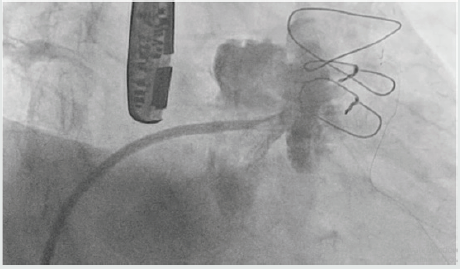
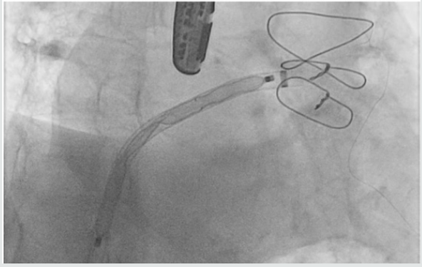
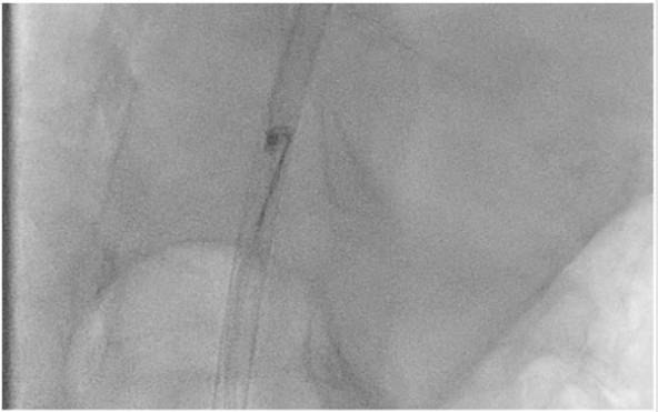
Lupine Publishers | Twine and Twine or Lose the Plug- Dislodged Left Atrial Appendage Closure Device
Lupine Publishers | Journal of Cardiology & Clinical Research
Abstract
Keywords: Amplatzer cardiac plug; Left atrial appendage occlusion; Device embolization
The following are the defined steps for ACP plug preparation:
a) Immerse device & hub end of loader in sterile saline to remove air.
b) Actively manipulate device in saline by hand to eliminate air bubbles on device.
c) Pull loading cable vice until lobe is fully retracted within loader, & stop before disc is recaptured
d) Insert distal end of delivery cable through haemostasis valve.
e) Connect delivery cable to exposed proximal end screw of device.
f) Grasp hub & rotate delivery cable clockwise until device is fully threaded on delivery cable.
g) Rotate delivery cable counter clockwise 1/8 of one turn to ensure that cable is not over tightened.
Numerous complications can be avoided if these steps are the done carefully and with utmost attention to every minute detail. In our case, we assume that excessive reverse rotation of the cable might be responsible for the device being loosely connected to the cable which got easily disconnected by the tug.
For
more Lupine Publishers Open
Access Journals Please visit our website
https://www.lupinepublishers.com/
For more Journal of Cardiology & Clinical Research Please Click Here: https://www.lupinepublishers.com/cardiology-journal/
For more Journal of Cardiology & Clinical Research Please Click Here: https://www.lupinepublishers.com/cardiology-journal/
Follow on Linkedin : https://www.linkedin.com/company/lupinepublishers
Follow on Twitter : https://twitter.com/lupine_online
Monday, September 23, 2019
Lupine Publishers: Lupine Publiahers | Radiocarpal Dislocation-a Comp...
Lupine Publishers: Lupine Publiahers | Radiocarpal Dislocation-a Comp...: Lupine Publishers | Journal of Orthoipaedics Abstract Go to The volar shearing distal radius fractures associated with subluxation...
Friday, September 20, 2019
Lupine Publishers Surgery Case Studies Journal: Lupine Publishers: Application of Geophysical and ...
Lupine Publishers Surgery Case Studies Journal: Lupine Publishers: Application of Geophysical and ...: Lupine Publishers: Application of Geophysical and Geotechnical Approa... : Lupine Publishers- Environmental and Soil Science Abstract ...
Lupine Publishers: Lupine Publishers Associate Editor Guidelines
Lupine Publishers: Lupine Publishers Associate Editor Guidelines: Lupine Publishers Associate Editor Guidelines Editorial Board members are chief support for our Lupine Publishers open access Journa...
Thursday, September 19, 2019
Lupine Publishers: Application of Geophysical and Geotechnical Approa...
Lupine Publishers: Application of Geophysical and Geotechnical Approa...: Lupine Publishers- Environmental and Soil Science Abstract Geophysical and Geotechnical investigations were carried out on the pr...
Wednesday, September 18, 2019
Lupine Publishers: The Biodiversity of Aquatic Gastropods in the Step...
Lupine Publishers: The Biodiversity of Aquatic Gastropods in the Step...: Lupine Publishers- Environmental and Soil Science Abstract This study describes the species diversity, abundance and...
Tuesday, September 17, 2019
Lupine Publishers: About Lupine Publishers
Lupine Publishers: About Lupine Publishers: About Lupine Publishers Lupine Publishers is one of the world’s largest open access publisher of peer-reviewed, fully peer reviewe...
Monday, September 16, 2019
Lupine Publishers: About Lupine Publishers
Lupine Publishers: About Lupine Publishers: About Lupine Publishers Lupine Publishers is one of the world’s largest open access publisher of peer-reviewed, fully peer reviewe...
Friday, September 13, 2019
Lupine Publishers: Lupine Publishers | A Combined Material Substituti...
Lupine Publishers: Lupine Publishers | A Combined Material Substituti...: Lupine Publishers | Journal of Textile and Fashion Designing Abstract This paper empirically presents a batik produ...
Wednesday, September 11, 2019
Lupine Publishers: Lupine Publishers | Green Revolution as Technologi...
Lupine Publishers: Lupine Publishers | Green Revolution as Technologi...: Lupine Publishers- Environmental and Soil Science Abstract The beginning of Human being’s effort to meet the need f...
Wednesday, September 4, 2019
Lupine Publishers: Lupine Publishers | Effect of Environment on Secon...
Lupine Publishers: Lupine Publishers | Effect of Environment on Secon...: Lupine Publishers- Environmental and Soil Science Abstract Medicinal plants constitute main resource base of almost all...
Tuesday, September 3, 2019
Lupine Publishers: Lupine Publishers | Forced Traction: An Error
Lupine Publishers: Lupine Publishers | Forced Traction: An Error: Lupine Publishers | Journal of Veterinary Science Introduction Immediate cause of dystocia requires certain preparations an...
Lupine Publishers: Lupine Publishers | Forced Traction: An Error
Lupine Publishers: Lupine Publishers | Forced Traction: An Error: Lupine Publishers | Journal of Veterinary Science Introduction Immediate cause of dystocia requires certain preparations an...
Subscribe to:
Posts (Atom)
Interruption of the Aortic Arch in the Adult and Fulminant Myocarditis: A Strange Presentation
Introduction 53 years old female patient, who presented oppressive precordial pain, radiating to the neck and jaw, for which she went to...


