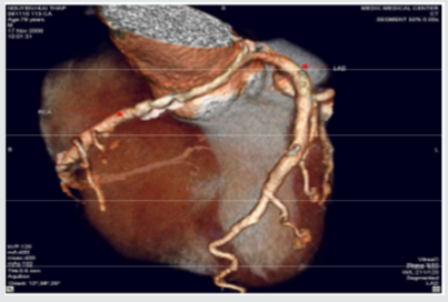
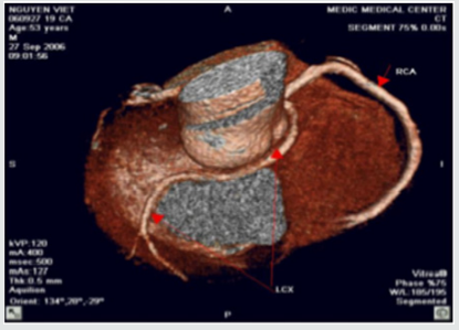
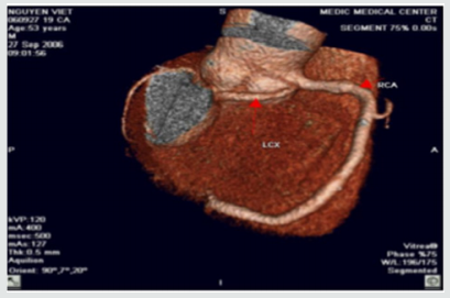
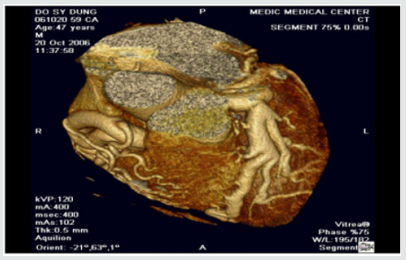
Showing posts with label Cardiovascular Research Journal. Show all posts

Friday, November 18, 2022

Profile of Patients admitted with Hypertension at a Tertiary Level Hospital of Eastern Nepal


Abstract

Hypertension is a global public health issue that affects approximately 1 billion people worldwide in both developed and developing countries. It is also considered as an ‘Iceberg’ disease’ because unknown morbidity far exceeds the known morbidity. High blood pressure (BP) is ranked as the third most important risk factor for at attributable burden of disease in South Asia. With an aim to determine the profile of the hypertensive patients a retrospective cross-sectional study design was used to analyze the medical record of 1311 patients admitted with the primary diagnosis of hypertension at BPKIHS January 2005 to May 2016. The study revealed that majority (82.4%) of the patients was above 40 years of age with the mean age of 57.14 ±17.5. More than half (53.5%) of the patients were male. Almost half (49.4%) of the patients were from Sun sari district. More than 90% of the patients were admitted in medical ward. Majority (90.7%) of the patients were improved at the time of discharge.

Read More About This Article Click on Below Link:
https://lupinepublishers.com/cardiology-journal/fulltext/profile-of-patients-admitted-with-hypertension-at-a-tertiary-level-hospital-of-eastern-nepal.ID.000102.php

Read More about Lupine Publishers Google Scholar Articles:
https://scholar.google.com/citations?view_op=view_citation&hl=en&user=EuBF0zMAAAAJ&citation_for_view=EuBF0zMAAAAJ:UY3hNwcQ290C

Saturday, June 26, 2021

Lupine Pub;oshers | Importance of Fast Brain MRI to confirm the Acute Stroke diagnosis after Thrombolysis

 Lupine Publishers | Advancements in Cardiology Research & Reports


Abstract

 Fast Brain MRI Protocol: Use of limited-sequence Brain MRI to confirm the diagnosis of stroke and identify ‘Stroke Mimics’ after negative initial neuroimaging following stroke thrombolysis.

Keywords: Stroke; Thrombolysis; Fast MRI (Magnetic resonance Imaging with limited sequence); CT-head (computerised Tomography of the head)

Introduction

Various studies have shown that a significant proportion of patients presenting as acute ischaemic stroke and thereby receiving stroke thrombolysis may have ‘Stroke Mimics’. Due to the time constrain in differentiating the true stroke from stroke mimics and limited availability of the timely MRI head these patients are not identified at stroke thrombolysis. However, diagnostic clarity is necessary thereafter for the appropriate management of such patients. A limited sequence ‘Fast Brain MRI protocol’ was introduced in our stroke unit to identify such ‘Stroke Mimics’ after thrombolysis. This 5-minute fast Brain MRI protocol included axial FLAIR and DWI sequence.

Methods

We evaluated all acute ischaemic stroke patients receiving thrombolysis in a DGH over 12 months. All thrombolysis patients received a routine CT Head 24 hours after receiving thrombolysis. Those patients with negative neuroimaging for an infarction at 24 hour received MRI Brain. A limited-sequence ‘Fast Brain MRI protocol’ was introduced in our stroke unit, this 5-minute fast Brain MRI protocol included axial FLAIR and DWI sequence[1].

Results

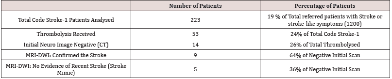
Out of total 1200 patents referred with a possible diagnosis of stroke or stroke-like event over the 12 months between August 2017 to July 2018, 223 patients were within the thrombolysis window. 53 of them received stroke thrombolysis (thrombolysis rate 24%)[2]. f 14 out of these 53 patients (26%) were neuroimaging negative at 24hour CT and hence ended up having Fast MRI of their head. Out of this 14 thrombolysed but initial neuroimaging-negative patients 6 patients (11% of total thrombolysed cases) were noted to have DWI-negative MRI Brain scan and hence identified as ‘Stroke Mimic’ who has received thrombolysis. Out of 14 thrombolysed but initial neuroimaging-negative cases 64% (9 out of 14) were confirmed to have a stroke after MRI scan and 36% (5 out of 14) were negative for any recent infarction (Table 1 & 2).

Table 1: Basic demography of the patients with acute stroke within the thrombolysis window.

Lupinepublishers-openaccess-cardiology

Table 2: Initial CT Head negative patients subsequent have fast Brain protocol MRI.

Lupinepublishers-openaccess-cardiology

Discussion

In our study, we noticed that the majority of patient with suspected acute ischaemic stroke and thereby receiving stroke thrombolysis were confirmed to have a cerebral infarction. Only 11% of thrombolysed stroke was confirmed as ‘Stroke mimic’. We concluded that thorough initial assessment and full NIHSS scoring had taken place in all these cases before thrombolysis. Unless a ‘Fast Brain MRI’ scan protocol is readily available without any delay prior to stroke thrombolysis it would not be possible to completely exclude the stroke, mimics receiving thrombolysis; as there is always anxiety that we might deny the thrombolysis to the genuine patients otherwise.

For more Lupine Publishers Click on Below link

https://publons.com/publisher/7295/lupine-publishers-llc


Saturday, June 19, 2021

Lupine Publishrers | Concomitant LVAD Implantation and Thoracic Surgery. Clinical Decision Making and Surgical Challenges

        Lupine Publishers | Advancements in Cardiology Research & Reports


Abstract

In the last years, left ventricular assist device (LVAD) indications have significantly broadened including candidates with multiple comorbidities even requiring combined surgical strategy. We present a case of a 56-year old patient affected by postischemic dilated cardiomyopathy in whom a lung nodule diagnosed during preoperative CT-scan could have contraindicated LVAD destination therapy. The patient underwent through a median sternotomy concomitant LVAD implantation on cardiopulmonary bypass followed by an atypical resection of the anterior part of the right lower lobe lung. A multidisciplinary, step-by-step approach to reduce the risk of right ventricular failure, bleeding and infections is presented.

Keywords: LVAD implantation; non-cardiac surgery; atypical lung resection

Indications to continuous flow left ventricular assist devices (CF-LVAD) have significantly broadened over the last decade considering even old patients with cardiac and extra-cardiac comorbidities [1]. Concomitant cardiac procedures to LVAD implantation are well described since nowadays up to 35% of implantations require concomitant cardiac surgery including valvular surgery, coronary artery bypass grafting, ventricular arrhythmias ablation, and atrial septal defects repair [1]. But the impact of these concomitant procedures is not well studied and guidelines are lacking. In particular for non-cardiac surgery experiences are limited to case series and the decision-making is driven on a single patient basis. To our knowledge, concomitant LVAD implantation and lung surgery has not been described before. We report the case of a patient who underwent a second generation CF-LVAD implantation followed by an atypical right lower lobe resection.

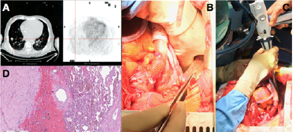
A 56-year-old man mildly obese and previous heavy smoker, presented with post-ischemic dilated cardiomyopathy and severely decompensated heart failure (orthopnea, dyspnea, fluid retention, and weight gain) with left ventricular ejection fraction (EF) of 17%. He also showed non-reversible post-capillary pulmonary hypertension and right ventricular (RV) dysfunction: Tricuspid Annular Plane Systolic Excursion (TAPSE) of 10 mm, RV Fractional Area Change (RVFAC) of 20%, Right Ventricular Stroke Work Indexed (RVSWI) of 400 mmHg/ml/m2 and a central venous pressure to wedge pressure ratio of 0.8. Few days after admission the patient developed hemoptysis. A 15-mm enhanced contrast lung nodule in the latero-basal segment of the right lower lobe (RLL) with a maximum standardized uptake value of 8, was demonstrated at the Positive Emission Tomography Scan (Figure 1A). The CT-scan showed a nodule suspicious for neuroendocrine tumor because of early contrast enhancement and a regular profile. Due to its peripheral position the nodule could not be reached with a transbronchial biopsy and a percutaneous computer tomography guided biopsy was considered to be too high risk. The pulmonary function tests were normal despite the presence of centrilobular and paraseptal emphysema.

Figure 1: (A)Computer Tomography and Positron Emission Tomography scan showing RLL enhanced contrast nodule; (B) manual palpation of lung nodule with deflated lung; (C) wedge resection of the RLL; (D) histology of the lung lesion showing a complex artero-venous malformation.


Lupinepublishers-openaccess-cardiology

He showed a positive clinical response to an aggressive diuretic therapy with significant reduction in weight and fluid retention, and improvement in right ventricular function (TAPSE 17 mm, RVFAC 33%, RVSWI 600 mmHg/mL/m2, central venous pressure to wedge pressure ratio of 0.4) which allowed to list the patient for an LVAD implantation. Considering right ventricular improvement, favorable position of the lung nodule and the complex coagulation management of a staged approach a combined procedure was preferred. A 24 hours infusion of levosimendan was completed in the immediate preoperative period. In consideration of the presence of the pulmonary lesion, we decided for a destination therapy configuration of the Jarvik 2000 (Jarvik Heart, Inc, New York, NY, USA) with an intraoperative retroauricolar implantation of the pedestal. After fixation of the pedestal, the pericardial space was approached via a median longitudinal sternotomy. Once the driveline was tunneled, LVAD implantation inside the left ventricle apex was performed on cardiopulmonary bypass (CPB) and beating heart, the outflow conduit was sutured to the ascending aorta. The progression from CPB to LVAD level 2 support was facilitated by continuous infusion of epinephrine (0.04 mcg/kg/min), norepinephrine (0.05 mcg/kg/min) and 20 ppm of inhaled nitric oxide to facilitate RV function.

When surgical and medical hemostasis were carefully achieved, one lung ventilation with 5 mL/Kg tidal volumes was initiated and a positive end expiratory pressure of 8 cmH2O, inhaled nitric oxide was increased to 30 ppm. Even if the PaO2/FiO2 ratio was 100 mmHg and the PaCO2 was 47 mmHg, the TEE showed normal RV function with a pulmonary artery systolic pressure of 40 mmHg. RLL exposure was achieved through the mediastinal pleura via the median sternotomic access. Manual palpation with inflated and noninflated lung allowed nodule identification and wedge resection of the RLL with an Echelon Flex 60 Endopatch (Ethicon Endo Surgery Inc., Cincinnati, OH) (Figure 1 B,C). Pathological examination demonstrated a complex artero-venous malformation (Figure 1D). The patient had an uneventful postoperative course with weaning from mechanical ventilation and inotropic support within the first 36 postoperative hours. He was discharged from the postoperative intensive care unit on day 6 and on day 17 to a rehabilitation center. Since the lung lesion was benign the patient was eligible to enter the heart transplantation list discharged and strictly monitored with a telemedicine program previously described [2].

The population of patients eligible for LVAD implantation has broadened over the last years and thanks to the reported improvements in mid-term outcomes and quality of life, the number of patients scheduled for a concomitant non-cardiac surgery intervention has progressively increased [3]. Planning a combined procedure in these fragile patients requires a thorough multidisciplinary approach, a strict preoperative medical optimization and the design of an effective and safe surgical procedure. Few cases of thoracic surgery in patients with CFLVAD have been published [4,5]. In this case, surgical plan was carefully defined in consideration of the major perioperative risks. Great attention was paid to RV function and preservation since hemodynamic changes following LVAD implantation are difficult to predict and have a huge impact on results [6,7]. Since no data have been published on the relationship between the amount of lung parenchyma excised and the variation of pulmonary resistances and thus of right ventricular afterload, we have chosen a step-bystep approach. An atypical lung resection was first performed. A RLL lobectomy would be a second surgical step only in case of demonstrated malignancy, minimizing in this way right ventricular impact and allowing a gradual adaptation of the RV to increased pulmonary resistances. During the procedure RV function was evaluated through surgical visual inspection, TEE and right heart catheterization allowing for immediate pump speed variations or pharmacological support titration.

The delicate interaction between RV, mechanical ventilation and hypoxia has been largely described in the population of patients suffering from acute respiratory distress syndrome and also during the intraoperative management of one lung ventilation for thoracic surgery, but it has never been studied in the context of LVAD support [8]. Indeed, RV disfunction after LVAD implantation is a life threating complication that can occur in 20 to 50% of patients [9] especially in the presence of high pulmonary vascular resistances. The RV showed a positive response to the increased venous return, to the geometrical adaptation of the interventricular septum and the augmented pulmonary vascular resistances. The favorable anatomical position of the lung nodule allowed to employ the median sternotomy for both LVAD implantation and lung resection. Concomitant procedure reduced the perioperative bleeding and infective risks. This case underlines the crucial role of a multidisciplinary approach, a preoperative medical optimization, a step-by-step surgical plan and a multimodal right ventricular evaluation.

For more Lupine Publishers Click on Below link

https://publons.com/publisher/7295/lupine-publishers-llc


Saturday, June 5, 2021

Lupine Publishers |A Comprehensive Overview of Risk Scoring Systems for Predicting Intravenous Immunoglobulin (IVIG)- Resistance in Kawasaki Disease

Lupine Publishers | Advancements in Cardiology Research & Reports


Abstract

It is important to predict Kawasaki Disease (KD) patients who will be resistant to Intravenous Immunoglobulin (IVIG) before starting the initial treatment, as these patients may have severe inflammation and vasculitis, which will likely lead to the development of Coronary Artery Lesions (CALs). An intensive initial treatment combined with IVIG and additional anti-inflammatory drugs is reported to reduce the occurrence of IVIG resistance and CALs. Although risk scoring systems using usual laboratory data to predict IVIG-resistant patients have mainly been developed in Japan, these systems did not accurately predict non-responders to IVIG among patients in the other countries. In this review, we provide a comprehensive overview of the main risk scoring systems and evaluate the relevant literature.

Kawasaki Disease (KD) is an acute systemic vasculitis that mainly occurs in infants and young children [1]. Although intravenous Immunoglobulin (IVIG) is an effective treatment for KD [2], approximately 10-20% of KD patients are resistant to IVIG therapy [2,3]. IVIG-resistant patients with KD have a higher risk of developing coronary artery lesions (CALs) than responders to IVIG therapy [4,5]. It is important to predict IVIG-resistant KD patients before starting the initial treatment, because intensive initial combination therapy with IVIG and other anti-inflammatory drugs, such as Ulinastatin [6], steroid [7,8] and infliximab [9], may reduce the occurrence of IVIG resistance and/or CALs. There are several risk scoring systems for predicting IVIG resistance in KD patients; the Kobayashi [10], Egami [11] and Sano [12] risk scores have been commonly used in Japan. Recently, we reported a new risk scoring system using two blood cell subtype ratios, the neutrophil-lymphocyte ratio (NLR) and the platelet-to-lymphocyte ratio (PLR) [13]. Furthermore, several researchers have reported other risk scoring systems in such countries as the U.S. [14], Taiwan [15] and China [16-18]. The aim of this review is to compare the predictive validity among these risk scoring systems and assess their problems and limitations.

The main risk scoring systems for predicting the IVIG resistance in KD, which have been reported to date, are summarized in Table 1. The parameters of the Egami score [11] consist of alanine aminotransferase (ALT) ≥80 IU/L (2 points), age ≤6 months (1 point), days of illness ≤4 days (1 point), C-reactive protein (CRP) ≥8 mg/dl (1 point) and platelet count ≤300×103/mm3 (1 point). In the high-risk group (score ≥3), the sensitivity and specificity in the prediction of IVIG resistance were 78% and 76%, respectively. The parameters of the Sano score [12] consist of Aspartate Amino Transferase (AST) ≥200 IU/L (1 point), CRP ≥7 mg/dl (1 point) and total bilirubin ≥0.9 mg/dl (1 point). In the high-risk group (score ≥2), the sensitivity and specificity in the prediction of IVIG resistance were 77% and 86%, respectively. The parameters of the Kobayashi score [10] consist of sodium ≤133 mmol/L (2 points), days of illness at initial treatment ≤4 days (2 points), AST ≥100 IU/L (2 points), % of neutrophils ≥80 (2 points), CRP ≥10 mg/dl (1 point), age ≤12 months (1 point) and platelet count ≤300×103/ mm3 (1 point). In the high-risk group (score ≥4), the sensitivity and specificity in the prediction of IVIG resistance were 86% and 68%, respectively. Recently, Kawamura et al. reported that the combination of NLR ≥3.83 and PLR ≥150 is a useful predictor of IVIG resistance in KD [13], and the sensitivity and specificity of NLR ≥3.83 and PLR ≥150 in the prediction of IVIG resistance were 71% and 69%, respectively. These simple ratios are convenient and costeffective in comparison to other scoring systems.

Table 1: Risk scoring systems predicting IVIG resistance in KD patients.


Lupinepublishers-openaccess-cardiology

ALT, alanine aminotransferase; AST, aspartate aminotransferase; CRP, C-reactive protein; GGT, γ-glutamyl transferase; NLR, neutrophil- lymphocyte ratio; PLR, platelet to lymphocyte ratio; PLT, platelet; zHgb, age-adjusted hemoglobin concentration.

In the US, the San Diego score [14] was proposed. The parameters consist of % of bands ≥20 (2 point), illness days≤4 (1 point), γ-glutamyl transferase (GGT) ≥60 IU/L and age-adjusted hemoglobin concentration (zHgb) ≤-2. In the high-risk group (score ≥2), the sensitivity and specificity in the prediction of IVIG resistance were 73% and 62%, respectively. In Taiwan, the Formosa score [15] was reported. The parameters consists of % of neutrophils ≥60 (2 points), albumin <3.5 g/dl (1 point) and positive lymphadenopathy (1 point). In the high-risk group (score ≥3), the sensitivity and specificity in the prediction of IVIG resistance were 86% and 81%, respectively. In China, Fu et al. reported a scoring system. The parameters consist of % of neutrophils ≥80 (2 points), illness days ≤4 (1 point), CRP ≥8 mg/dl (2 pint), polymorphous exanthema (1 point) and change around the anus (1 point) [16]. In the high-risk group (score ≥4), the sensitivity and specificity in the prediction of IVIG resistance were 54% and 71%, respectively. Tang et al. reported another scoring system. The parameters consist of age <6 months (2 points), albumin 3.5 < g/dl (2 points), edema of extremities (1 point), rash (1 point) and % of neutrophils ≥80 (1 point) [17]. In the high-risk group (score ≥3), the sensitivity and specificity in the prediction of IVIG resistance were 71% and 76%, respectively. Recently, Hua et al. reported a new scoring system. The parameters consist of fever duration ≥7 days (2 points), delayed diagnosis (1 point), GGP ≥25 mg/dl (1 point), sodium < 135 mmol/L (1 point), NLR ≥2.8 (1 point) and platelet count ≤350×103/mm3 (1 point) [18]. In the high-risk group (score ≥4), the sensitivity and specificity in the prediction of IVIG resistance were 61% and 67%, respectively. As described above, each of risk scoring systems are determined based on different clinical data and symptoms, although some factors are duplicated among these scoring systems.

There are differences in the definition of IVIG resistance in each study. Egami defined IVIG resistance as persistent fever (≥37.5℃) and a fall in CRP by <50% within 48 hours after IVIG therapy [11]. Sano defined IVIG resistance as persistent fever (≥37.5℃ over 24 hours) after finishing IVIG therapy [12]. Kobayashi and Kawamura defined IVIG resistance as persistent fever lasting >24 hours after the completion of the initial treatment or in the presence of recrudescent fever associated with KD symptoms after an afebrile period [10,13]. The San Diego score defined IVIG resistance as persistent fever (≥38.0℃ rectally or orally) for at least 48 hours but no longer than 7 days after IVIG therapy [14]. The Formosa score defined IVIG resistance as persistent fever or development of recrudescent fever associated with KD symptoms after afebrile period [15]. Fu and Hua defined IVIG resistance as persistent or recrudescent fever at any time 48 hours to 2 weeks after IVIG therapy and at least 1 of the standard diagnostic criteria [16,18]. Tang defined IVIG resistance as recrudescent or persistent fever ≥36 hours after the end of IVIG infusion [17]. Thus, because the definition of IVIG resistance has not been standardized, international consensus will be needed in the near future. In the 2017 Kawasaki disease guidelines from the American Heart Association, the definition of IVIG resistance was recrudescent or persistent fever at least 36 hours after the end of IVIG infusion [19].

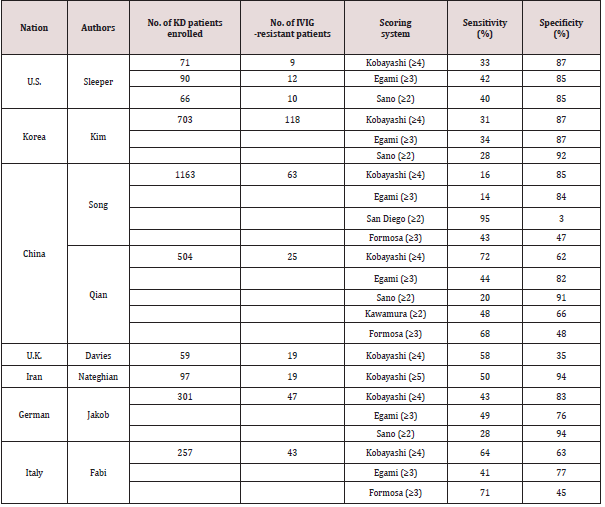
Several authors have assessed the sensitivity and specificity of risk scoring systems when they were applied to KD patients in the other countries (Table 2). The Kobayashi risk score (≥4), Egami risk score (≥3) and Sano risk score (≥2) have good specificity (87%, 85% and 85%, respectively) but low sensitivity (33%, 42% and 40%, respectively) for predicting IVIG resistance in KD patients in North America [20]. Similarly, the Kobayashi risk score (≥4), Egami risk score (≥3) and Sano risk score (≥2) have good specificity (87%, 87% and 92%, respectively) but low sensitivity (31%, 34% and 28%, respectively) for predicting IVIG resistance in KD patients in Korea [21]. In KD patients in China, Song et al. reported that the Kobayashi risk score (≥4) and Egami risk score (≥3) have good specificity (85% and 84%, respectively) but low sensitivity (16% and 14%, respectively), the San Diego risk score (≥2) has high sensitivity (95%) but very low sensitivity (3%), and the Formosa score (≥3) has relatively low specificity (47%) and sensitivity (43%) for predicting IVIG resistance [22]. Qian et al. reported that the sensitivity of Kobayashi risk score (≥4), Egami risk score (≥3), Sano risk score (≥2), Kawamura risk score (≥2) and Formosa score (≥3) were 72%, 44%, 20%, 48% and 68%, respectively, and that the specificity of these scores were 62%, 82%, 91%, 66% and 48%, respectively [23]. In the United Kingdom, the Kobayashi risk score (≥4) had relatively low sensitivity (58%) and low specificity (35%) [24]. In the Kobayashi score, a cut-off risk score of 5 points was also reported to be effective for predicting IVIG resistance in Japanese patients with KD [7,25]. The Kobayashi risk score (≥5) is reported to predict IVIG resistance in Iranian patients with KD, with 50% sensitivity and 94% specificity [26].

Table 2: Sensitivity and Specificity of risk scoring systems when applied to different ethnic group.


Lupinepublishers-openaccess-cardiology

Recently, Jakob et al. reported that the Kobayashi risk score (≥4), Egami risk score (≥3) and Sano risk score (≥2) have low sensitivity (43%, 49% and 28%, respectively), although they have relatively high specificity (83%, 76% and 94%, respectively), in German patients with KD [27]. More recently, Fabi et el. reported that the Kobayashi risk score (≥4), Egami risk score (≥3) and Formosa score (≥3) are ineffective for predicting IVIG resistance (sensitivity: 64%, 41% and 71%, respectively; specificity: 63%, 77% and 45%, respectively) in Italian children with KD [28]. Besides, the ability of NRL and PLR to predict IVIG resistance in KD was evaluated in China: the cut-off values of NLR ≥4.36 and PLR ≥162 were useful for predicting IVIG-resistance in KD [29], and NLR ≥2.51 was useful in KD patients younger than 1 year of age [30]. Although there is a slight difference in the cut-off values of Japan [13] and China [29], the effectiveness of the NLR and PLR in predicting IVIG resistance has been proven in both countries.

Many of the Japanese scoring systems (Egami, Sano and Kobayashi scores) had relatively good specificity but low sensitivity when they were applied to non-Japanese KD patients. These results indicate that the use of Japanese risk scores in other countries can exclude most patients who do not require additional therapy (lowrisk patients) but cannot accurately extract patients who require additional therapies (high-risk patients). For this reason, these Japanese risk scores have not been widely used outside Japan. These regional differences could be due to genetic differences or other environmental factors [31]. It is reported that the functional polymorphism and methylation of the immunoglobulin gamma Fc region receptor II-a (FCGR2A) gene might be associated with IVIG resistance in KD patients [32,33]. As there is a difference in the incidence of KD among countries, the disease severity and the effectiveness of IVIG therapy might also be different. It seems difficult to establish a universal risk scoring system for IVIG resistance in KD due to racial differences. Thus, it might be better to aim to establish discrete risk scoring systems for each country. It would be preferable if the risk score is simple and convenient. The determination of cut-off values for the NLR and PLR in each country may warrant investigation because these ratios are easily calculated. In summary, the prediction of failure to respond to IVIG therapy is important for identifying KD patients who may need additional anti-inflammatory treatments, because intensive therapy can be reduce the incidence of IVIG resistance and CAL formation. Although several risk scoring systems of IVIG resistance have been proposed, many of these failed to effectively predict IVIG resistance in other countries. Further studies will be needed to obtain consensus on a risk scoring system for predicting IVIG resistance in KD.


For more Lupine Publishers Click on Below link

https://publons.com/publisher/7295/lupine-publishers-llc


Friday, February 12, 2021

Lupine Publishers | Epidemiological Aspects of Cardiac Decompensation Factors Renaissance Hospital N’Djamena Chad

Lupine Publishers | Advancements in Cardiology Research & Reports


Abstract


Introduction: Cardiac decompensation factors are numerous. Their identification allows better management of patients and limits the rate of rehospitalization. The aim of this work was to identify cardiac decompensation factors and improve their management at the renaissance hospital in N’Djamena, Chad.

Patients and Methods: This was a cross-sectional retrospective study conducted in the cardiology department at N’Djamena Renaissance Hospital, over a period of one year, from 01 January 2018 to 01 January 2019. All patients hospitalized for cardiac decompensation during this period and consented, were included.

Results: During the period of our study, 52 patients were included. The sex ratio was 1.9. The mean age was 48±9 years old. The predominant cardiovascular risk factors were arterial hypertension (37%, n = 19) and diabetes (27%, n = 14). The monthly income of our patients was in the majority of cases less than 200,000 FCFA (44%, n = 23). The main factors of cardiac decompensation were respectively, infections (18%, n = 9), supraventricular arrhythmias (16%, n = 8), changes in temperature (11%, n = 6), therapeutic nonobservance (11%, n = 6), dietary gap (10%, n = 5), and hypertensive relapses (10%, n = 5). The main etiologies of heart failure were ischemic cardiomyopathies (31%, n = 16), dilated cardiomyopathies (25%, n = 13), hypertensive cardiomyopathies (17%, n = 9), and rheumatic valvulopathies (15%). %, n = 8).

Conclusion: Rehospitalizations for cardiac decompensation are common after the first episode of hospitalization. The identification of the risk factors for this decompensation and their management make it possible to avoid these readmissions.

Keywords: Cardiac decompensation factors; Heart failure; Renaissance hospital N’Djamena characterized

Abbreviations: HF: Heart Failure; FFCA: Franc of the Financial Community in Africa; AFA: Atrial Fibrillation Arrhythmia; LV: Left Ventricle

Introduction

Heart failure (HF) is defined as a clinical syndrome characterized by chronic symptoms (dyspnea, fatigue) that may be accompanied by physical signs (crepitus, peripheral edema) caused by a structural cardiac abnormality and / or functional, resulting in decreased cardiac output [1]. It is a major public health problem because of its frequency and consequences in terms of morbidity and mortality and its economic impact on the health care system. Its prevalence is increasing because of the aging of the population but also because of the improvement of the management of many heart diseases including ischemic heart disease. It is one of the leading causes of hospitalization, morbidity and mortality, especially among the elderly [2]. Its evolution is clinically marked by periods of remission and exacerbation leading to recurrent hospitalizations. The number of readmissions for IC remains significant despite the therapeutic progress of recent years. The identification of decompensation factors and the optimization of their management could prevent these readmissions, particularly after hospitalization. The objective of this study was to identify cardiac decompensation factors and improve their management at the renal hospital in N’Djamena, Chad.

Patients and Methods

This was a cross-sectional retrospective study conducted in the cardiology department at N’Djamena Renaissance Hospital, over a period of one year, from 01 January 2018 to 01 January 2019. Were included all patients readmitted for cardiac decompensation during the study period and who gave their consent.

The parameters studied

Epidemiological characteristics: age, sex, cardiovascular risk factors (arterial hypertension, diabetes, chronic renal dysfunction with glomerular filtration rate <60ml/min / 1.73m2, obesity, dyslipidemia, alcohol, smoking), monthly cost of treatment in FFCA (1 US dollar = 593,720 FFCA). Clinical features: cardiac decompensatory factors (difference in diet, unsuitable exercise, temperature, alcohol, AFA, other rhythm disorders, hypertensive pressure, ischemic episode, anemia, bronchopulmonary infection, other infection, renal failure, poor compliance drug, hyperthyroidism, untreated sleep apnea syndrome), etiologies of HF. Electrocardiographic characteristics: arrhythmias (atrial or ventricular extrasystoles, atrial fibrillation or flutter, atrial tachycardia, ventricular tachycardia), repolarization abnormalities, sinoatrial or atrioventricular blocks. Echocardiographic features: dilated cardiac cavities, wall hypertrophy, diastolic dysfunction of the left ventricle (LV); abnormalities of left ventricular kinetics (hyperkinesia, hypokinesia, akinesia), LV systolic dysfunction (systolic ejection fraction <45%), valvular abnormality, congenital anomaly, pulmonary arterial hypertension.

Statistical Analysis

In this study, a descriptive statistical analysis was applied using Microsoft Excel, quantitative variables were presented by their mean and standard deviation and qualitative variables were by percentages.

Ethics

This work was done by obtaining the approval of the hospital management and the consent of the patients.

Results

During the period of our study, 52 patients were included. Men predominated with 65% of cases (n = 34). The sex ratio was 1.9. The average age was 48±9 years old with a minimum age of 22 years and a maximum age of 79 years. Patients were educated only in 38% of cases (n = 20). The predominant cardiovascular risk factors were hypertension (19%), diabetes (14%), dyslipidemia (11%), and 19%). The monthly income of our patients was in the majority of cases less than 200,000 FCFA. The rates were, respectively, between CFAF 100,000 and 200,000 in 44% of cases (n =23) and less than CFAF 100,000 in 33% of cases (n = 17). Only 12 patients (23%) had an income above 200,000 FCFA. The health insurance rate was observed in 13% of cases (n=7). (Table 1) shows the characteristics of the patients. The main factors of cardiac decompensation are shown in Table 2. The most frequent were respectively, infections in 18% of cases (n = 9), including 4 cases of bronchopulmonary infections (8%), supraventricular rhythm disorders in 16% of cases (n=8) including 5 cases of AFA (10%), changes in temperature especially heat with 6 cases (11%), poor therapeutic compliance 6 cases (11%), the difference diet in 10% of cases (n = 5), and hypertensive outbreaks in 10% of cases (n=5). The most common etiologies of IC (Table 3) were ischemic cardiomyopathies in 31% of cases (n=16), of which 4 patients (8%) had benefited from myocardial revascularization, dilated cardiomyopathies in 25% (n=16). = 13), hypertensive cardiomyopathies in 17% of cases (n =9) and rheumatic valvulopathies in 15% (n=8). Other etiologies were less frequent in this series such as post-embolic pulmonary heart in 6% of cases (n=3), congenital heart disease in 4% (n=2) and pericardial affections in 2% of cases (n= 1).

Table 1: Patient Characteristics.

Lupinepublishers-openaccess-cardiology

Table 2: Cardiac Decompensation Factors.

Lupinepublishers-openaccess-cardiology

Table 3: Etiologies of HF.

Lupinepublishers-openaccess-cardiology

Discussion

The post-hospitalization period is conducive to rehospitalization, but long-term chronic HF patient follow-up is important because the morbidity and mortality rate remains high in this group of patients, even though many treatments have been shown to be effective. The follow-up of these patients makes it possible to optimize the therapeutics, the monitoring and to detect early the signs of decompensation. Cardiac rehabilitation plays an important role in the management of HF and should be part of the modern strategy for the management of patients with stable heart failure. This includes not only physical training, but also rehabilitation of drug treatments, control of risk factors, psychological management and finally, patient education [3]. The search for this factor that shifts a situation of balance during a decompensation is fundamental because its treatment can be very profitable. Several standards for HF support highlight the need to look for a triggering factor [4]. We studied the importance of triggers for cardiac decompensation in a population of readmitted patients who were followed for chronic HF. The main triggering factors identified were, in order of frequency, infections, atrial fibrillation, therapeutic nonobservance, temperature variations, mainly heat, uncontrolled hypertension, diet deviation, anemia, myocardial ischemia. These factors are globally found in the literature data. In this study, the first triggering factor found was the existence of an infection (18%) with bronchopulmonary infections (8%) at their head. They are promoted by decreasing the effectiveness of coughing, bronchiolar elasticity, efficiency of the mucociliary system and swallowing disorders.

The mechanisms involved in cardiac decompensation are multiple. Several studies have highlighted the important role of infections, particularly bronchopulmonary infections, in cardiac decompensation, especially in elderly patients [5-8]. The second triggering factor was a supraventricular rhythm disorder (16%), especially ACFA (10%). Suppression of atrial systole results in increased LV filling pressures and promotes the onset of HF thrust. AFA is common in HF and its prevalence increases with the severity of CI [9-13]. Cardiac decompensation was attributed to noncompliance in 8% of the cases in this study, but adherence remains difficult to assess. This non-compliance was favored by several factors in our patients; the high number of drugs with their adverse effects, given the high rate of comorbidities observed (diabetes, high blood pressure, kidney failure), the economic level which was low in almost 50% of patients and the absence of health insurance, the level of education and the influence that might have on the understanding of the disease. The therapeutic nonobservance in the HF varies in the literature from 10 to 99% according to the evaluation method used [14]. Compliance is responsible for an increase in the number of hospitalizations and a worsening of clinical signs [15]. A multidisciplinary intervention has shown its effectiveness on adherence to 30-day treatment [16] and an educational intervention has improved compliance and decreased re-hospitalization rates [17]. Anemia was found in 4 patients in our series.

This result is weak compared to data from the literature because, according to several authors, anemia is frequent during IC and is a poor prognostic factor in chronic HF [18,19], increased clinical signs, aggravation of functional status [20]. However, there is little data on the involvement of acute anemia in cardiac decompensation. In addition, the management of the etiology of HF remains essential to prevent complications and readmissions and to improve the prognosis. In our study we found that in coronary patients (31%) only 8% had benefited from myocardial revascularization, valvular patients (15%) and those with congenital heart disease (4%) had not received reparations. surgical procedures that were necessary. In our context, the identification of the decompensation factors must be of paramount importance for the clinician and should allow the improvement of the management of the pathology in particular at the preventive level. Hence the importance of educating the patient and those around him. The establishment of appropriate structures and care networks for heart failure in all regions of Chad will allow better monitoring of patients with rapid access to the specialist physician.

The limits of our work

Our study presents several methodological limitations. First, it is a retrospective study with information gathered from the reports that sometimes did not contain all the necessary data. The size of our sample is small, we will have to lead other more representative studies in the future. And many patients have not received etiologic treatment of HF.

Conclusion

Rehospitalizations for cardiac decompensation are common after the first episode of hospitalization. The identification of the risk factors for this decompensation and their management make it possible to avoid these readmissions. Hence the importance of emphasizing access to care with appropriate therapeutic means, regular monitoring and therapeutic education.

For more Lupine Publishers Click on Below link

https://www.zoominfo.com/c/lupine-publishers-llc/43353514


Friday, January 22, 2021

Lupine Publishers | Relationship between Depression and Physical Activity of Myocardial Infarction Patients after Treatment

Lupine Publishers | Advancements in Cardiology Research & Reports


Abstract


Physical activity is a secondary prevention that can reduce mortality and re-admission in patients with myocardial infarction. The objective of this study was to identify the relationship with physical activity of myocardial infarction patients after treatment. This study used a cross sectional method. A total of 150 myocardial infarction patients were selected using a purposive sampling technique. The results showed that the majority of STEMI post-treatment patients have mild physical activity (82%). There is also a significant relationship between depression and the level of physical activity of myocardial infarction patients after treatment (p = 0.003), OR = 0.144 (95% CI; 0.032-0.635). Depression in myocardial infarction patients at the time of the attack, if not intervened properly, it will persist and affect physical activity after treatment. A recommendation is directed to the nursing department to assess depression in patients with newly diagnosed of myocardial infarction.

Keywords: Physical activity; myocardial infarction; depression in myocardial infarction patients

Background

Physical activity is recommended by the European Society of Cardiology (ESC) as a long-term therapy in prevention for myocardial infarction patients Ibanez et al. [1]; Amsterdam et al. [2]. Physical activity can reduce mortality, re-admission and improve the quality of life of patients with myocardial infarction Andersen & Laustsen, [3]; Dalal, et al. [4]; Ek et al. [5]. Although physical activity is recommended as a long-term therapy in STEMI patients, only 37% of patients actively engage in physical activity after treatment Mckee et al. [6]. Several factors are known to have an association with physical activity in myocardial infarction patients, one of which is depression Mckee et al. [6]. Depression in myocardial infarction patients occurs 48-72 hours after a heart attack Kala, et al. [7]. Depression has a negative effect on post-treatment recovery in myocardial infarction patients, and causes lower compliance to treatment programs Homma et al. [8]; Kumar et al. [9]. The objective of this study was to identify the relationship between depression and physical activity of patients with myocardial infarction after treatment.

Method

This design of the study was a cross sectional study. Sampling was carried out using non-probability sampling techniques with a sample of 150 people. The inclusion criteria in this study were patients aged ≥18 years who were diagnosed with myocardial infarction. The study was conducted at the regional hospital of Jambi province, Indonesia in February - March 2018. Data collection was done using PHQ-9 Patient Depression Questionairre Kroenke et al. [10] and International Physical Activity Questionnaire (IPAQ) Strath et al. [11].

Findings

Characteristics of Respondents

Of the 150 post-treatment STEMI patients, the majority of patients were aged 18-60 years (73.3%), were male (78.7%), and 75.3% of whom had passed 7 to 30 days post-treatment. The majority of respondents were in the category of mild depression 69.3%, whereas the rest experienced moderate-severe depression, and 82% of respondents were at the level of physical activity with mild categories. Relationship between depression and physical activity of post-treatment myocardial infarction patients. The results of the analysis of the relationship between depression and physical activity showed that 95.7% of post-treatment myocardial infarction patients experienced moderate-severe depression with mild physical activity. Meanwhile, among post-treatment myocardial infarction patients who experience mild depression, 24% had moderate-heavy physical activity. Fisher exact test results obtained p = 0.003, so it can be concluded that there is a relationship between depression and physical activity. From the results of the analysis also demonstrated that the value of OR is 0.144 (95% CI; 0.032-0.635). By looking at the OR values it can be concluded that post-treatment myocardial infarction patients who experience mild depression would have a 0.144 times greater chance of having moderate-heavy physical activity compared to patients who have moderate-severe depression category

Discussion

Physical activity is a key component in heart disease patients that is beneficial in reducing the risk of relapse Thompson et al. [12]. In this study, the results of the analysis showed that 82% of respondents are at the level of mild physical activity. The results of this study are similar to studies conducted by Matthias, 2017 in Sri Lanka, where 56, 7% of respondents had low physical activity Matthias et al. [9]. Low physical activity is a trigger for the occurrence of myocardial infarction. Physical activity increases the process of arteriosclerosis formation, decreases inflammation, and triggers the formation of thrombosis Cheng et al. [13]. Many factors can affect physical activity. The study of Mckee et al. [6]. concluded that depression was one of the dominant factors causing low physical activity. This is the same as the results of this study. The results of bivariate testing found a relationship between depression and physical activity. Patients with myocardial infarction who experience depression tend to smoke, have low physical activity, and consume a lot of alcohol Qing Wu et al. [14]. In addition, experience during an attack is a cause of depression, and this continues for up to two months after the attack. This state of depression results in the patient becoming silent and limiting their physical activity.

Conclusion

Post-treatment myocardial infarction patients have a mild level of physical activity, and depression during the attack still occurs in myocardial infarction patients after undergoing treatment in the hospital. Depression, if not properly intervened, will cause changes in physical activity of myocardial infarction patients after treatment.

For more Lupine Publishers Click on Below link

https://www.zoominfo.com/c/lupine-publishers-llc/433535148

Monday, January 11, 2021

Lupine Publishers | MDCT in Diagnosis of Anomalies of Coronary Artery Origin and Course a Coronary MDCT-Angiographic study of 9572 patients

Lupine Publishers | Advancements in Cardiology Research & Reports


Abstract

Background: Coronary anomalies are the causes of sudden cardiac deaths in young peoples, but usually asymptomatic. We perform this retrospective study to determine the types and prevalence of Coronary Anomalies of origin and course.

Method: The data of 9572 patients with Coronary CT-angiography by MDCT 640 Aquilion Toshiba machine were analyzed.

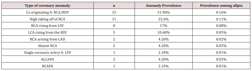
Results: Anomalous origin and course of coronary artery were detected in 47 (0.49%) of 9572 patients. The anomalous origins of Circumflex Artery from the RCA or the right sinus of Valsalva are most frequently visualized ( 15 pts [31.9%] ). High taking off of RCA observed in 11 pts ( 23.4% ).The RCA rising from the left sinus of Valsalva were seen in 8 pts ( 17% ).The Left Coronary Artery originates from the right sinus of Valsalva in 5 pts ( 10,6% ).The RCA arising from the LAD in 2pts (4,2% ).Absent RCA in 2 case (4.2%) and single coronary artery from LSV in one case (2.1%). The LCA rising from the Pulmonary Artery ( ALCAPA) in 2 cases and The RCA originating from the PA in one case ( RCAPA ).

Conclusion: Anomalies of coronary artery origin and course are rare but the diagnosis is very important to prevent SCD in young patients. MDCT with the Volume Rendered Images is the non-invasive modality that provides the valuable information to detect these anomalies.

Keywords: Multidetector Computed Tomography; Anomalies of coronary origin and course; sinus of Valsalva

Introduction

Coronary artery anomalies are a diverse group of congenital heart diseases with manifestations and pathological mechanisms are highly variable. Coronary anomalies include anomalies of origin and course, anomalies of intrinsic coronary arterial anatomy like myocardial bridge, anatomy of coronary termination as coronary artery fistula and anomalous anastomotic vessels. Anomalies of coronary origin and course may associated with arrhythmias, myocardial infarction and sudden cardiac deaths in young people, especially on effort like athletes. We study 9572 patients with coronary MDCT-angiography to evaluate the type and the incidence of coronary anomalies of origin and course[1,2].

Methods

All patients who underwent coronary CT-angiography by MDCT 64O Aquilion Toshiba equipment ( IV contrast medium, gantry rotation of 0.33 msec, slice thickness 0.5mm ) in MEDIC HCMC Viet Nam, from January 2016 to January 2019 were included. The main indications of CT-angiography were acute coronary syndrome, stable angina, coronary CT-angiography prior to surgery, congenital heart diseases involving coronary artery...

The CT-angiograms with coronary anomalies were selected and analyzed. The anomalies of coronary origin and course were assessed [3-5].

Results

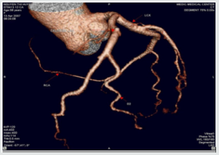
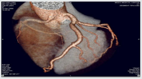
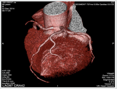
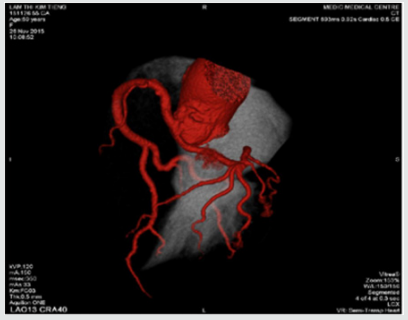
We included 9572 pts with anomalies of coronary origin and course based on results of CT-angiograms that were interpreted by two cardiologists. Anomalous origin and course of coronary artery were detected in 47 ( 0,49 %) of 9572 patients. The mean age of these pts was 63± 8.4, M/F=1.8 . The anomalous origins of Circumflex Artery from the RCA or the right sinus of Valsalva are most frequently visualized ( 15 pts [31.9%] ).High taking off of RCA observed in 11 pts ( 23.4% ) The RCA rising from the left sinus of Valsalva were seen in 8 pts ( 17% ).The Left Coronary Artery originates from the right sinus of Valsalva in 5 pts ( 10.6% ), in this subgroup, a patient presented by myocardial infarction resulting cardiac arrest was notified, the surgical re-implantation of LCA was performed .The RCA arising from the LAD in 2pts (4,2% ). Absent RCA in 2 case (4.2%) and single coronary artery from LSV in one case ( 2.1% ) (Table1 ).The Left Coronary Artery arising from the Pulmonary Artery ( ALCAPA ) in 2 cases ( 4.2% ) and The RCA originating from the PA ( RCAPA ) in one case ( 2.1% ). sinus of Valsalva (Figures 1-10).

Table 1.

Lupinepublishers-openaccess-cardiology

RSV: Right sinus of Valsalva, LSV: Left sinus of Valsalva, ALCAPA: Anomalous Left Coronary Artery from The Pulmonary Artery, RCAPA: Anomalous Origin of the Right Coronary Artery off The Pulmonary Artery.

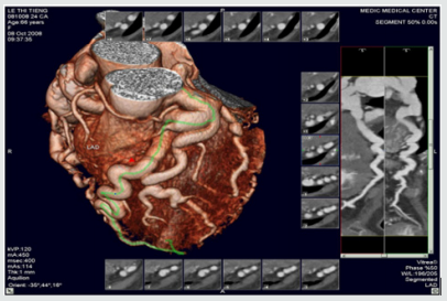
Figure 1: Single coronary artery rising from LSV.

Lupinepublishers-openaccess-cardiology

This patient is of 52 ages, presented by atypical chest pain, the single coronary artery originating from LSV. The other case report of Prashanth Panduranga revealed the single coronary artery arising from RSV with exertional angina

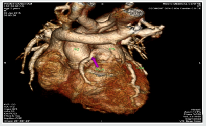
Figure 2: High taking off of RCA.

Lupinepublishers-openaccess-cardiology

Some time causes myocardial infarction due to excessive angulation between RCA and Aorta. We have in our study one young patient of 24 y.o that had been transferred to the hospital by cardiac arrest , related to this anomaly. Operative re-implanted had been indicated to save the patient

Figure 3: RCA originates from LSV with intra-arterial course resulting Angina

Lupinepublishers-openaccess-cardiology

Figure 4: Anomalous origin of LCA from RSV

Lupinepublishers-openaccess-cardiology

Figure 5: RCA rising from LSV and Intra-arterial course of RCA.

Lupinepublishers-openaccess-cardiology

Figure 6:LCx arising from the RVS and Retro Aortic Course of LCx.

Lupinepublishers-openaccess-cardiology

Figure 7: LCx arising from the RVS and Retro Aortic Course of LCx.

Lupinepublishers-openaccess-cardiology

Figure 8: Anomalous Left Coronary Artery from The Pulmonary Artery.

Lupinepublishers-openaccess-cardiology

Figure 9: Other case of ALCAPA.

Lupinepublishers-openaccess-cardiology

Figure 10: Anomalous Origin of the Right Coronary Artery off The Pulmonary Artery.

Lupinepublishers-openaccess-cardiology

Discussion & Conclusion

In our study, coronary anomalies of origin and course were detected in 47 of 9572 patients ( 0,49% ) that is consistent with the incidence of 0.27% to 1.66% reported in other series. The most frequent anomaly of origin and course was the Cx Artery arising from RCA/RSV ( 31.9% of anomaly prevalence and 0.16% among all patients ), this incidence is lower than previous published studies. The anomalies of origin and course of RCA were found in 17% and 4.2% respectively from LSV and LAD. This incidence is lower in comparison with previous study. Sudden deaths, myocardial infarction, arrhythmias related to the coronary anomalies were reported previously [6,7]. But these anomalies often asymptomatic, so early detection of coronary anomalies of origin and course is highly important. The former studies mainly based on the result of coronary angiography that is invasive modality. This study demonstrates MDCT is the noninvasive modality that provides important information related to coronary anatomy. Currently MDCT and MRI become fundamental to detection and diagnosis of coronary anomalies. Contrast enhanced ECG-gated 640-row MDCT coronary angiography is an accurate diagnostic method that can precisely detect the coronary anomalies of origin and course.


For more Lupine Publishers Click on Below link

https://www.zoominfo.com/c/lupine-publishers-llc/433535148


Friday, December 11, 2020

Lupine Publishers | Platelets and Patent Ductus Arteriosus: Is there a Any Association Really?

Lupine Publishers | Advancements in Cardiology Research & Reports


Abstract


Introduction: A great number of the studies have shown that platelets play a role in closure of the PDA. However, studies that reported that platelet parameters were not associated with PDA were also published. We also wanted to contribute to clarify the relationship between PDA and platelet parameters.

Materials and Methods: Preterm infants that less than 34 gestational weeks were examined to echocardiography at the time of detected clinical findings or within 24-72 h after admission to our unit, routinely. The patients were divided into two groups according to echocardiography findings randomly; hsPDA require ductal closure treatment and non-hsPDA. The platelet count, MPV, PDW, PCT and Platelet Mass Index values of both groups were compared.

Results: There was no difference between the two groups in terms of MPV, Platelet count and Platelet mass index. However, PDW and PCT were statistically significantly in the study group than the control group.

Discussion: As a result, according to our study, platelet count, MPV and platelet mass index cannot be used to predict either hsPDA or treatment success, but a low PCT and high PDW can be used predict hsPDA but not treatment success.

Patent Ductus Arteriosus (PDA) can cause mortality and morbidity such as respiratory distress syndrome (RDS), pulmonary hemorrhage, bronchopulmonary dysplasia (BPD), intraventricular hemorrhage (IVH), necrotizing enterocolitis (NEC), retinopathy of prematurity (ROP) [1]. For this reason, early diagnosis and treatment of PDA is the most important point. The main diagnostic method of PDA is Doppler echocardiography [2]. However, there is no clearly consensus on diagnosis of hemodynamically significant patent ductus arteriosus (hsPDA). Therefore, new diagnostic methods of PDA are needed. A great number of the studies have shown that platelets play a role in closure of the PDA [3-6]. The first of these studies, Echtler et al. studied the relationship between ductal closure and platelet parameters in animals [3]. In the same study, the ductus arteriosus did not close (thus, remained permanently open) in animals in which platelet functions were compromised. After this study, they studied on premature infants about relationship between ductal closure and platelet parameters. According to this study, a low platelet count and a low PDW were risk factors for PDA. However, studies that reported that platelet parameters were not associated with PDA were also published [7-11]. We also wanted to contribute to clarify the relationship between PDA and platelet parameters.

This observational, retrospective cohort study was conducted between August 2017 and 2018. Preterm infants that less than 34 gestational weeks were examined to echocardiography at the time of detected clinical findings or within 24-72 h after admission to our unit, routinely [12]. The patients were divided into two groups according to echocardiography findings randomly; hsPDA require ductal closure treatment and non-hsPDA. hsPDA was defined when at least one of the clinical findings associated with PDA was present: a hyperdynamic precordium; a sustained murmur; tachycardia; hypotension; oliguria; an increased pulse pressure; an increase in ventilation pressure and/or oxygen demand; and at least one echocardiographic finding: ductal diameter ≥1.5mm, left atrium/ aortic root ratio ≥1.5, and/or diastolic flow failure in the abdominal aorta or inverse flow. We applied intravenous or oral ibuprofen to close the hsPDA. Intravenous or oral paracetamol was given in cases who ibuprofen is unsuccessful or contraindicated. After treatment, echocardiography was performed again, and the PDA was classified as open or closed. We excluded those with conditions that might cause inflammation or affect platelet count and/or function (Antenatal steroid use, PPROM, early sepsis, chorioamnionitis, congenital viral infections, preeclampsia), congenital heart disease, pulmonary hypertension, perinatal asphyxia, congenital anomaly, chromosomal anomaly, thrombocytopenia (<50.000/mm3), and lack of data. Written informed consent was obtained from all parents. All echocardiographic examinations were performed by Vivid S6 Echocardiography System fitted with a 10S transducer (General Electric Healthcare, Milwaukee, WI, USA). Blood samples taken from an umbilical venous catheter at between 48-72 hours and 7. day, were collected in ethylenediaminetetraacetic acid-containing tubes and blood counts performed using a Coulter Counter model LH (Coulter Electronics, Hialeah, FL, USA). This yielded the platelet count, MPV, PDW, PCT. The platelet mass index was obtained from the platelet count (103/mm3) and the MPV (fL). We recorded gestational age, birth weight, sex, mode of delivery, Apgar scores (at 1 and 5 min) 48-72 h and 7. day platelet parameters, surfactant requirement, ventilation history, IVH, NEC, ROP, BPD, duration of hospitalization and any death.

Statistical analyses were performed using SPSS for Windows ver. 22.0 (SPSS Inc., Chicago, Illinois). The paired samples t-test and independent samples t-test were used to compare continuous variables. Continuous variables are presented as means ± SDs, and categorical variables are given as frequencies with percentages. A p-value less than 0.05 was considered statistically significant.

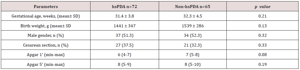
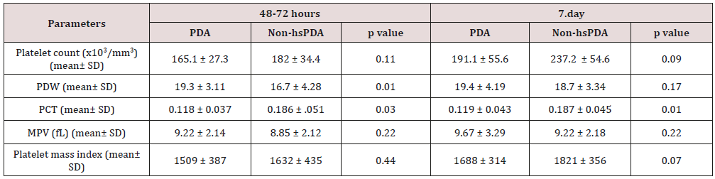
258 newborns under 34 weeks were admitted to our unit, of whom 121 were excluded. The study group consisted of 72 premature infants with hsDPA who applied ductus closure treatment and 65 premature infants without hs DPA or spontaneously closed PDA consisted of the control group (Figure 1). The demographic characteristics of both groups are shown in Table 1. The mean gestational age and the mean birth weight of the study and control groups were, respectively, 31.4±3.8 vs. 32.3±4.5 weeks (p=0.12); 1441±347 vs. 1,539±286g (p=0.08). There was no difference between two groups in perinatal parameters. The platelet parameters of both groups are shown in Table 2. There was no difference between the two groups in terms of MPV, Platelet count and Platelet mass index. However, PDW and PCT were statistically significantly in the study group than the control group.

Table 1: Comparison perinatal characteristics of the study and control groups.


Lupinepublishers-openaccess-cardiology

hsPDA: hemodynamically significant patent ductus arteriosus.

Table 2: Comparison of the platelet parameters of the study and control groups.


Lupinepublishers-openaccess-cardiology

PDW: platelet distribution width; PCT: platocrit; MPV: mean platelet volume; Platelet mass index: the platelet count (103/mm3) X MPV (fL); hsPDA: hemodynamically significant patent ductus arteriosus.

Figure 1: Flowchart of study.


Lupinepublishers-openaccess-cardiology

PPROM: Preterm premature rupture of the membranes; hsPDA: hemodynamically significant patent ductus arteriosus.

Low oxygen pressure, elevated prostaglandin and nitric oxide levels are the main factors affecting continuity of the ductus arteriosus in the uterus. After birth, increased oxygen levels and decreased prostaglandin levels enable functional closure of the DA [13]. In addition to this mechanism, different mechanisms of closure of the ductus began to be discussed. The discussion began when Echtler et al. showed that platelets were attached to the lumen of the closed ductus arteriosus and confirmed this experimental finding via a retrospective study of preterm births [3]. After this animal study, various hypotheses about the role played by platelets in duct closure in newborns have been proposed. The most acceptable hypothesis is an effect of platelets on DA contraction, which occurs immediately after birth in term newborns, triggering hypoxia in the vessel wall by decreasing the blood flow in the venous lumen and vasa vasorum; in preterm newborns, the cells in the ductus wall are fed by the ductal lumen because of the absence of a vasa vasorum. As the ductus wall is thin, contraction is inadequate and endothelial damage and platelet aggregation thus develop because of vesselwall hypoxia. Based on this hypothesis, it was claimed that platelet counts were important in terms of DA closure in preterm infants, as they are in the pathophysiology of adult vascular diseases [14,15]. However, this hypothesis is not supported by the fact that platelet transfusion does not reduce the incidence of PDA in preterm newborns with immune thrombocytopenia and does not increase the PDA frequency in term newborns with severe thrombocytopenia secondary to Wiskott-Aldrich syndrome [16-20].

In Fujioka et al. [21-23]. the platelet count was not related to PDA diagnosis or treatment success. On the other hand, Echtler et al. [3,5,6] reported that a low platelet count increased the hsPDA incidence [24-25]. In some works performed after these contradictory studies, it was reported that large platelets create a greater potential risk of prothrombotic reactions; large platelets are more aggregated than small and normal platelets given the greater number of receptors such as thromboxane A2-B2 and glycoproteins IIb-IIIa on the surfaces of large platelets. It was suggested that the increased metabolic and enzymatic activities of dysfunctional thrombocytes, rather than the platelet count, were associated with PDA [26-29]. We sought to identify parameters related to platelet function associated with PDA. These remain controversial; all of MPV, PDW, PCT, and platelet mass index have been associated with cardiovascular diseases in adults [30-35]. In addition, in a limited number of studies on neonates, the MPV and PDW were shown to be associated with prematurity complications such as RDS and BPD [36-37].

In our study, no difference was found between the platelet counts of the hsPDA and control groups at 48-72 h and 7. day. In addition, there was no difference between the platelet counts of newborn who did and did not fail treatment. In conclusion, the platelet count was not a predictor of hsPDA diagnosis or treatment success. The results of our study contradict those of the two major meta-analyses conducted by Simon et al. and Mitra et al. but support the cohort study of Sallmon et al. [18-20]. PCT was lower and PDW was higher in the study groups than control groups and the difference between the two groups was statistically significant. However, MPV and platelet mass index were similar in both groups. Thus, we conclude that the PCT and PDW can be used to predict hsPDA but not treatment success. Demirel and Dizdar et al. [4]. reported that the PDW was higher in preterm infants with hsPDA than in control groups [38,39]. Bekmez et al [40]. reported that a low PCT increased the hsPDA incidence Demir et al.[41]. found a high MPV and a low platelet mass in the hsPDA group. We also excluded patients who received ibuprofen as ductus closure therapy because of potential effects on platelet count and functions. Infants born to mothers with prior pre-eclampsia, which affects platelet count and ductal flow because of the increased placental resistance, were also excluded [42-44]. We also excluded infants with perinatal asphyxia associated with an increased PDA, thrombocytopenia, and platelet dysfunction [45-47]. Newborns whose mothers had earlier received steroids were excluded because of possible effects on the platelet count. We thus excluded all pathologies that may affect platelet count and function and induce inflammation. There were some limitations of our study. The first limitation of our study is that it was retrospective in nature. The second limitation is modest sample size. As a result, according to our study, platelet count, MPV and platelet mass index cannot be used to predict either hsPDA or treatment success, but a low PCT and high PDW can be used predict hsPDA but not treatment success.

For more Lupine Publishers Social Bookmarking Click on Below link

https://issuu.com/lupinepublishers-oaj/docs/lupine_publishers_social_bookmarking_sites.docx


Interruption of the Aortic Arch in the Adult and Fulminant Myocarditis: A Strange Presentation

Introduction   53 years old female patient, who presented oppressive precordial pain, radiating to the neck and jaw, for which she went to...