Showing posts with label Cardilogy. Show all posts

Monday, May 3, 2021

Lupine Publishers | Blinded by the Common Causes: Ischemic Monomelic Neuropathy; a Rare Complication after Vascular Access Creation

Lupine Publishers | Advancements in Cardiology Research & Reports


Abstract

The authors discuss a case of a rare complications of vascular access creation in a hemodialysis patient “ischemic monomelic neuropathy”. This is an ischemic axonal injury to the nerves supplying the left hand after arterio-venous graft placement for hemodialysis. The authors also discuss the diagnosis, pathophysiology and treatment of this rare vascular access complications that sometimes occurs in hemodialysis patients immediately following arterio-venous fistula or graft (AVF or AVG) placement.

Keywords: Ischemic Monomelic Neuropathy; Arterio-Venous Fistula or Graft; Hemodialysis; Nerve Conduction Studies; Steal Syndrome

Case History

The patient is 73-year Hispanic male with past medical history of hypothyroidism, type-2 diabetes mellitus, hypertension, hyperlipidemia, and uremic encephalopathy diagnosed recently for which he was started on hemodialysis on May 2019. He had a left upper arm loop graft placed on June,13 2019, immediately after the surgery the patient complained of pain, numbness and tingling of the fingers and swelling of the of the left hand. He failed to followup with his surgeon. In the meantime, his symptoms got worse with increasing pain and swelling followed by loss of function of the left hand to the point that he could not grip objects with his hand.

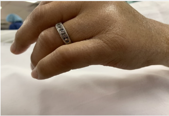
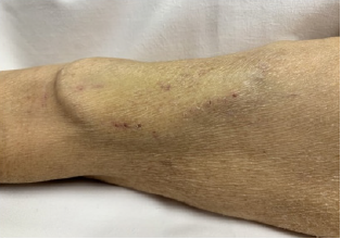
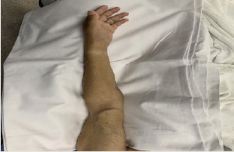
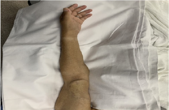
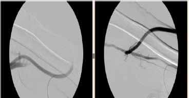
He was referred to the Surgery and Vascular Center at Regency Park, Toledo, Ohio on the 8/28/2019 for fistulogram with a diagnosis of vascular steal syndrome. Clinical examination showed a thin elderly man in no acute distress. His vital signs were within normal limits. Examination of the cardio-vascular and respiratory systems were normal. He has a loop graft in the left upper arm with good thrill and bruits. His radial and ulnar pulses were palpable and within normal limits. The patient was not able to use the hand and fingers to grip objects or squeeze the examiner’s hand. He has loss of sensation over the distributions of the medial and ulnar nerves with swelling of the left hand (Figures 1-5). The angiogram of the graft was normal (Figure 6). A preliminary diagnosis of ischemic monomelic neuropathy was entertained based on the clinical picture and the normal angiogram of the access. He was referred for nerve conduction studies and surgical consultation for possible ligature of the graft.

Figure 1:
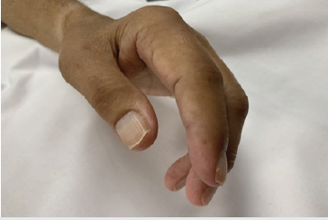
Lupinepublishers-openaccess-cardiology

Figure 2:

Lupinepublishers-openaccess-cardiology

Figure 3:

Lupinepublishers-openaccess-cardiology

Figure 4:

Lupinepublishers-openaccess-cardiology

Figure 5:

Lupinepublishers-openaccess-cardiology

Figure 6:

Lupinepublishers-openaccess-cardiology

Case Discussion

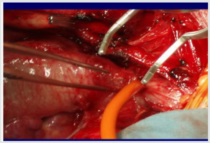
Ischemic monomelic neuropathy (IMN) is a combined sensory and motor impairment without major vascular or tissue necrosis. It is a form of ischemia of the nerves that supply the upper limbs especially the hand. It occurs immediately after vascular access creation for hemodialysis patients. It is a rare complication and caused by ischemic axonal loss of the nerves that supply the distal arms [1-3]. IMN is first reported by Wilbourn et al in the year 1983 [1]. IMN is under-recognized and mis-diagnosed, but its known incidence is 0.5 to 3% after vascular creations according to recent reports [3]. “What the mind cannot conceive the eyes cannot see”. If a hemodialysis patient complains of hand pain after placement of arterio-venous fistula or graft, the physician needs to consider many diseases, including soft tissue swelling, wound hematoma, carpal tunnel syndrome, vascular steal syndrome, and IMN [4-6]. The most important factor in the diagnosis of IMN is to think of it and correlate and interpret the symptoms, signs of the clinical examination of the access and rule out vascular steal syndrome. Acute pain, weakness, and muscle paralysis immediately after AVF/AVG placement are common warning signs and symptoms for the presence of IMN. Since these symptoms are non-specific, after vascular access creation, the motor and sensory function of the operated hand should be examined carefully, and nerve conduction studies should be carried out urgently. Low amplitudes and reduced or even undetectable motor or sensory nerve conduction velocities in the presence of preservation of the vascular integrities of the hand are compatible with IMN. Axonal degeneration of the median, radial, and ulnar nerves can be observed [3].

Electromyogram (EMG) often show degeneration, including fibrillation potentials and motor unit loss. Past neuropathy, diabetes mellitus, atherosclerosis, upper arm vascular access, and female gender have increased risk for IMN [4,7-9]. These factors compromise the brachial artery which is the only blood supply to the distal arm. The most effective treatment of IMN is early recognition of the condition and immediate closure of the access; this increase the probability of recovery [1,4]. Early closure of the vascular access leads to partial or full recovery of the sensory and motor function of the hand [8]. The easiest way is to tie up the fistula or graft as soon as possible [5]. Better awareness and education of the surgeons and nephrologists should lead to early diagnosis and proper management of this dreadful complication (IMN) [4]. So, educating our minds to conceive the rare complications of vascular access surgeries are the best way to train the eyes to easily identify the complications before it is too late to help our patients.


For more Lupine Publishers Click on Below link

https://publons.com/publisher/7295/lupine-publishers-llc

Friday, January 22, 2021

Lupine Publishers | Relationship between Depression and Physical Activity of Myocardial Infarction Patients after Treatment

Lupine Publishers | Advancements in Cardiology Research & Reports


Abstract


Physical activity is a secondary prevention that can reduce mortality and re-admission in patients with myocardial infarction. The objective of this study was to identify the relationship with physical activity of myocardial infarction patients after treatment. This study used a cross sectional method. A total of 150 myocardial infarction patients were selected using a purposive sampling technique. The results showed that the majority of STEMI post-treatment patients have mild physical activity (82%). There is also a significant relationship between depression and the level of physical activity of myocardial infarction patients after treatment (p = 0.003), OR = 0.144 (95% CI; 0.032-0.635). Depression in myocardial infarction patients at the time of the attack, if not intervened properly, it will persist and affect physical activity after treatment. A recommendation is directed to the nursing department to assess depression in patients with newly diagnosed of myocardial infarction.

Keywords: Physical activity; myocardial infarction; depression in myocardial infarction patients

Background

Physical activity is recommended by the European Society of Cardiology (ESC) as a long-term therapy in prevention for myocardial infarction patients Ibanez et al. [1]; Amsterdam et al. [2]. Physical activity can reduce mortality, re-admission and improve the quality of life of patients with myocardial infarction Andersen & Laustsen, [3]; Dalal, et al. [4]; Ek et al. [5]. Although physical activity is recommended as a long-term therapy in STEMI patients, only 37% of patients actively engage in physical activity after treatment Mckee et al. [6]. Several factors are known to have an association with physical activity in myocardial infarction patients, one of which is depression Mckee et al. [6]. Depression in myocardial infarction patients occurs 48-72 hours after a heart attack Kala, et al. [7]. Depression has a negative effect on post-treatment recovery in myocardial infarction patients, and causes lower compliance to treatment programs Homma et al. [8]; Kumar et al. [9]. The objective of this study was to identify the relationship between depression and physical activity of patients with myocardial infarction after treatment.

Method

This design of the study was a cross sectional study. Sampling was carried out using non-probability sampling techniques with a sample of 150 people. The inclusion criteria in this study were patients aged ≥18 years who were diagnosed with myocardial infarction. The study was conducted at the regional hospital of Jambi province, Indonesia in February - March 2018. Data collection was done using PHQ-9 Patient Depression Questionairre Kroenke et al. [10] and International Physical Activity Questionnaire (IPAQ) Strath et al. [11].

Findings

Characteristics of Respondents

Of the 150 post-treatment STEMI patients, the majority of patients were aged 18-60 years (73.3%), were male (78.7%), and 75.3% of whom had passed 7 to 30 days post-treatment. The majority of respondents were in the category of mild depression 69.3%, whereas the rest experienced moderate-severe depression, and 82% of respondents were at the level of physical activity with mild categories. Relationship between depression and physical activity of post-treatment myocardial infarction patients. The results of the analysis of the relationship between depression and physical activity showed that 95.7% of post-treatment myocardial infarction patients experienced moderate-severe depression with mild physical activity. Meanwhile, among post-treatment myocardial infarction patients who experience mild depression, 24% had moderate-heavy physical activity. Fisher exact test results obtained p = 0.003, so it can be concluded that there is a relationship between depression and physical activity. From the results of the analysis also demonstrated that the value of OR is 0.144 (95% CI; 0.032-0.635). By looking at the OR values it can be concluded that post-treatment myocardial infarction patients who experience mild depression would have a 0.144 times greater chance of having moderate-heavy physical activity compared to patients who have moderate-severe depression category

Discussion

Physical activity is a key component in heart disease patients that is beneficial in reducing the risk of relapse Thompson et al. [12]. In this study, the results of the analysis showed that 82% of respondents are at the level of mild physical activity. The results of this study are similar to studies conducted by Matthias, 2017 in Sri Lanka, where 56, 7% of respondents had low physical activity Matthias et al. [9]. Low physical activity is a trigger for the occurrence of myocardial infarction. Physical activity increases the process of arteriosclerosis formation, decreases inflammation, and triggers the formation of thrombosis Cheng et al. [13]. Many factors can affect physical activity. The study of Mckee et al. [6]. concluded that depression was one of the dominant factors causing low physical activity. This is the same as the results of this study. The results of bivariate testing found a relationship between depression and physical activity. Patients with myocardial infarction who experience depression tend to smoke, have low physical activity, and consume a lot of alcohol Qing Wu et al. [14]. In addition, experience during an attack is a cause of depression, and this continues for up to two months after the attack. This state of depression results in the patient becoming silent and limiting their physical activity.

Conclusion

Post-treatment myocardial infarction patients have a mild level of physical activity, and depression during the attack still occurs in myocardial infarction patients after undergoing treatment in the hospital. Depression, if not properly intervened, will cause changes in physical activity of myocardial infarction patients after treatment.

For more Lupine Publishers Click on Below link

https://www.zoominfo.com/c/lupine-publishers-llc/433535148

Wednesday, December 23, 2020

Lupine Publishers | Msct In Diagnosis of Congenital Heart Diseases in Viet Nam

Lupine Publishers | Advancements in Cardiology Research & Reports


Abstract

Background: Congenital heart diseases associated with more malformations, complex aortopulmonary collaterals and anomalous coronary artery. Echocardiography is the initial diagnostic method but this method can be limited in complex congenital heart diseases.
Purpose: To assess the role of MDCT in congenital heart diseases (CHD) diagnosis compare with operative result and interventional angiography.

Methods: 910 patients with congenital heart diseases of 31.000 patients underwent cardiac angiography with 64 and 320 section CT at Medic Medical Center since 09/09/2006 to 30/12/2015.

Results: There are 658 operated cases, most of operated cases demonstrated the exact diagnosis of MDCT in congenital heart diseases.

Conclusions: MDCT is the fast and non-invasive diagnostic method with the high accuracy, overcomes the limit of echocardiography in complex congenital heart diseases diagnosis and provides the panorama and useful information’s prior to the operation.

Keywords: Congenital heart diseases; Cardiac multi-detector computed tomography, Multi-detector computed tomography in congenital heart diseases; Congenital heart diseases computed tomography

Introduction

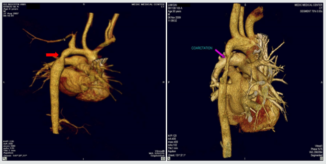
Congenital heart diseases effect ~ 1% of all live births in the general population. Complex congenital heart diseases associated with more malformations, complex aortopulmonary collaterals and anomalous coronary artery. Over the past few decades, the diagnosis and treatment of congenital heart diseases have greatly improved [1-6]. Diagnostic tools: X-ray, ECG, echocardiography, MRI and MDCT. ECG and X-Ray suggest the diagnosis but are not specific. Echocardiography is the initial diagnostic method for patients with suspected CHD but this method can be limited in complex CHD. The great capabilities of MRI for anatomic and functional assessment of the heart but MRI is time-consuming and may require patient sedation. Now enable CT to be used as an accurate noninvasive clinical instrument that is fast replacing invasive cine-angiography in the evaluation of CHD [1,2,5].

I. Improves both spatial and temporal resolution.

II. Increases scanning speed.

III. Improves diagnostic image quality by reducing respiratory artifacts

Purpose

To assess the role of MDCT in congenital heart diseases (CHD) diagnosis compare with operative result and interventional angiography.

Material and Methods

Subject: 910 patients with congenital heart diseases of 31.000 patients underwent cardiac angiography with 64 and 640 section CT at Medic Medical Center since 09/09/2006 to 30/12/2015.

Means and scanning techniques

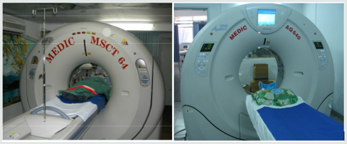
a) Medic Medical Center scanned cardiac CT by 64 MDCT Toshiba Aquilion machine and Toshiba Aquilion One (320 MDCT), 0.5mm slice thickness, 0.5mm imaging reconstruction.

b) Two phases scanning: Don’t inject phase and contrast media injection phase: +Phase doesn’t inject contrast which help locate and assess coronary artery calcification.

c) +Phase inject contrast media: Medicine chasing phase and water chasing phase.

d) Contrast pumping machine is double-barreled Stellant (Medrad).

e) To inject contrast by intravenous right hand.

f) Contrast dose used 1mL/ kg.

g) Drug pump speed depends on patient status and disease.

h) Vitrea software: Reconstructed images by MPR, MIP and VRT.

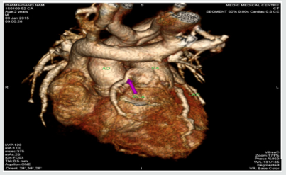
i) Effective radiation dose is low (320-MDCT is 3.69±061mSv; 64-MDCT is 12-14mSv) (Figures 1).

Figure 1.

Lupinepublishers-openaccess-cardiology

Data analysis

a. The prospective study and case series report compare with operative and interventional angiography.

b. Data collection at the HCM city Heart Institute, Tam Duc Heart Hospital and Medic medical center (Figures 2-17).

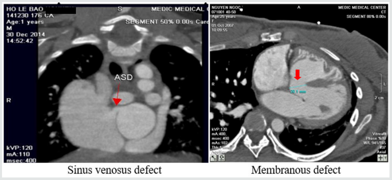
Figure 2: Atrial septal defects and Ventricular septal defects.

Lupinepublishers-openaccess-cardiology

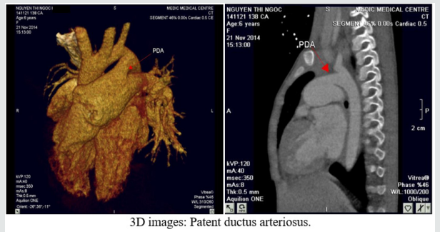
Figure 3: Patent ductus arteriosus.

Lupinepublishers-openaccess-cardiology

Figure 4: Coarctation of aorta.

Lupinepublishers-openaccess-cardiology

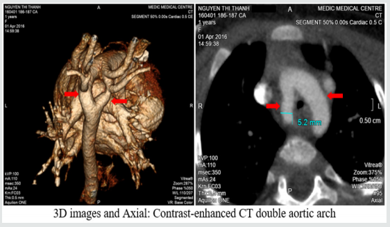
Figure 5: Double aortic arch.

Lupinepublishers-openaccess-cardiology

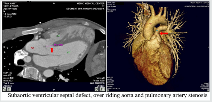
Figure 6: Tetralogy of Fallot.

Lupinepublishers-openaccess-cardiology

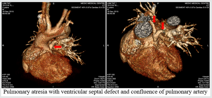
Figure 7: Pulmonary atresia with ventricular septal defect.

Lupinepublishers-openaccess-cardiology

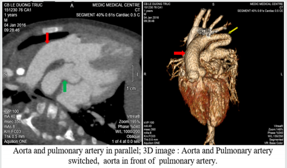
Figure 8: Transposition of great vessels.

Lupinepublishers-openaccess-cardiology

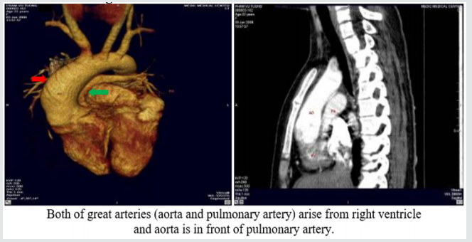
Figure 9: Double outlet right ventricle.

Lupinepublishers-openaccess-cardiology

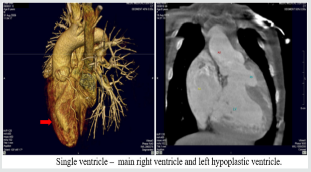
Figure 10: Single ventricle.

Lupinepublishers-openaccess-cardiology

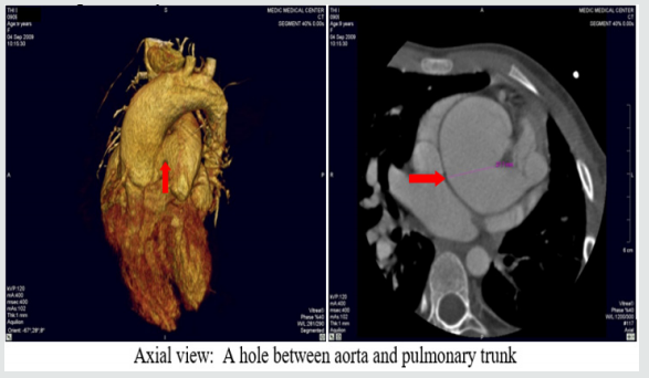
Figure 11: Aortopulmonary window:

Lupinepublishers-openaccess-cardiology

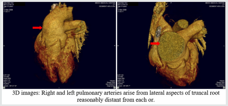
Figure 12: Truncus arteriosus.

Lupinepublishers-openaccess-cardiology

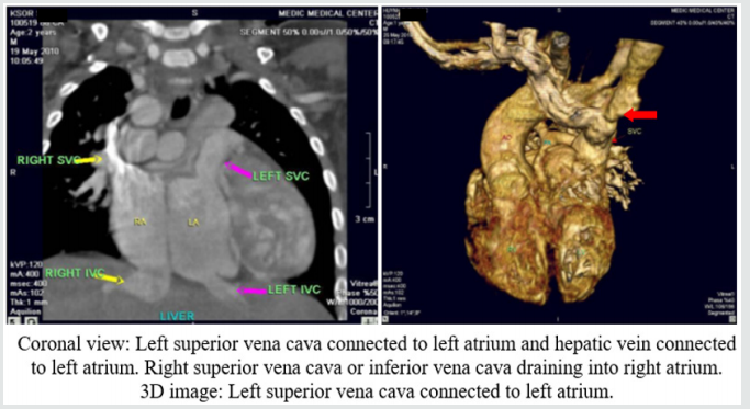
Figure 13: Anomalous systemic venous return.

Lupinepublishers-openaccess-cardiology

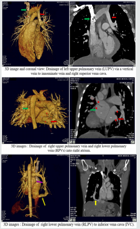
Figure 14: Anomalous pulmonary venous connection.

Lupinepublishers-openaccess-cardiology

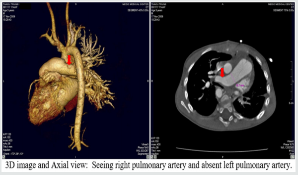
Figure 15: Single pulmonary artery.

Lupinepublishers-openaccess-cardiology

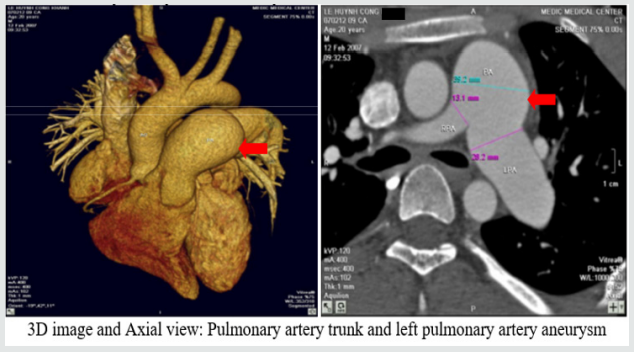
Figure 16: Pulmonary artery trunk aneurysm:

Lupinepublishers-openaccess-cardiology

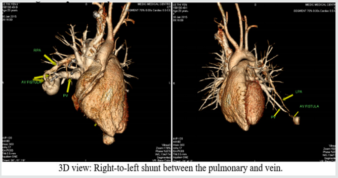
Figure 17: Congenital pulmonary arteriovenous malformation.

Lupinepublishers-openaccess-cardiology

Results

There are 658 operated cases, most of operated cases demonstrated the exact diagnosis of MDCT in congenital heart diseases.

Discussion

Congenital heart diseases associated with more malformations, complex aortopulmonary collaterals and anomalous coronary artery. Echocardiography is the initial evaluative method for preand post-operation congenital heart diseases but this method can be limited in complex cases. Multi-detector computed tomography overcomes the limit of Echocardiography by multiplanar reconstruction (MPR) and volume rendered techniques (VRT) reconstruction . Volume rendered techniques (VRT) reconstruction clearly demonstrates the relationship between the heart and great vessels.

Conclusion

Multi-detector computed tomography is the fast and noninvasive diagnostic method with the high accuracy. Overcomes the limit of Echocardiography in complex congenital heart diseases. Provides the panorama and useful information’s prior to the surgery.

For more Lupine Publishers Social Bookmarking Click on Below link

https://issuu.com/lupinepublishers-oaj/docs/lupine_publishers_social_bookmarking_sites.doc


Friday, December 18, 2020

Lupine Publishers | The Left Common Carotid Artery Rises from the Main Pulmonary Artery

Lupine Publishers | Advancements in Cardiology Research & Reports


Abstract

A young female patient of 15y.o presented at my hospital by dyspnea on effort and palpitation for one year. Mental deficiency was notified. Physical examination detected a 3/6 continuous murmur at the 2ndRICS. In the past history, PDA had been suspected by her physician, associated with recurrent bronchitis. Trans-thoracic Echocardiography showed an enlarged LV of 57mm with normal EF of 69% , LCA=5mm, RCA=3.5mm at origin, no suspected sign of PDA was seen. Only a continuous flow was visualized in the PA. CT-Angiography with IV contrast medium showed the Left Common Carotid Artery rising from the Pulmonary Artery trunk. PDA was not presented. The Left Common Carotid Artery then was re-implanted into the aortic arch normally with a favorable postoperative progress.

Keywords: Carotid Artery; Pulmonary Artery; Anomalous origin

Anomalous origin of the left common carotid artery is very rare and has been reported previously. We present an operated case of this topic with clinical finding, cardiac ultrasound and MDCT imaging.

Figure 1: Right aortic Arch.


Lupinepublishers-openaccess-cardiology

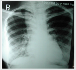
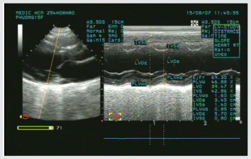
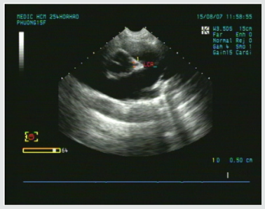
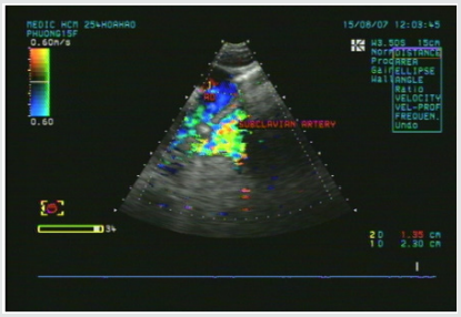
A young female patient of 15y.o presented at my hospital by dyspnea and palpitation when running and fast walking for one year. Mental deficiency was notified, she had some difficulties to learn at school. Physical examination detected a 3/6 continuous murmur at the 2ndRICS. In the past history, PDA has been suspected by her physician, associated with recurrent bronchitis. Her body state was normal with 1m60 of height and 48 kg of weight. She was evaluated immediately by a chest X ray that showed a right aortic arch (Figure 1). The trans-thoracic echocardiography that revealed an enlarged LV of 57mm with normal EF of 69% (Figure 2), LCA=5mm, RCA=3.5mm at origin (Figure 3). No suspected sign of PDA was detected except a continuous flow presented in the Pulmonary Artery (Figure 4).

Figure 2: Enlarged LV& normal systolic function.


Lupinepublishers-openaccess-cardiology

Figure 3: Normal LCA at origin.


Lupinepublishers-openaccess-cardiology

Figure 4: Continuous flow in the PA.


Lupinepublishers-openaccess-cardiology

Figure 5: Absence of aortic origin of the LCCA.


Lupinepublishers-openaccess-cardiology

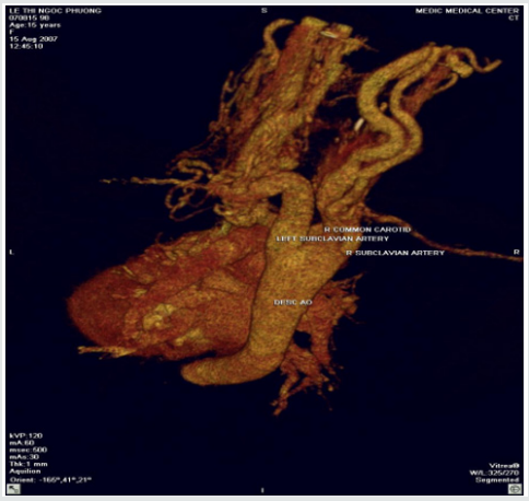
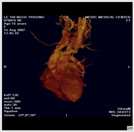
CT-Angiography (MDCT 64) with IV contrast medium Ultravist, slice thickness=1mm visualized a right aortic arch, aberrant origin of the left subclavian artery, dilatation of the branches rising from aortic arch with increased collateral vessels (Figure 5). Especially, MDCT 64 showed the Left Common Carotid Artery ( LCCA ) rose from the PA trunk (Figure 6) PDA was not detected. Patient underwent uncomplicated surgical repair: the Left Common Carotid Artery was re-implanted into the aortic arch normally with a favorable post-operative progress (Figure 7).

Figure 6: The LCCA rising from the Main PA roof.


Lupinepublishers-openaccess-cardiology

Figure 7: Re-implantation of the LCCA.


Lupinepublishers-openaccess-cardiology

Anomalous origin of the Left Common Carotid Artery from the Pulmonary Artery Trunk has been previously reported as rare cases. Kagami Mijaji et al. [1] has reported a case of anomalous origin of the Artery from the Right Pulmonary Artery. Onyekachukwu et al. [2] has described a case of anomalous origin of the Left Common Carotid Artery from the Main Pulmonary Artery. In this article, my patient was not infant with CHARGES syndrome that includes multiple congenital anomalies like the patients in their reports. She was a teenage patient without other congenital disease. The role of ultrasound is orienting for the indication of Computed Tomography or DSA. In case of present turbulent flow in the PA, Coronary Fistula and other shunts from the head and neck vessels should be considered [3].

Anomalous origin of the Left Common Carotid Artery is very rare congenital defect that maybe isolated or associated with some syndromes. Noninvasive diagnostic methods as Ultrasound and CTA may confirm the diagnosis and inform the anatomical relation of the anomalous vessels prior to operate.

For more Lupine Publishers Social Bookmarking Click on Below link

https://issuu.com/lupinepublishers-oaj/docs/lupine_publishers_social_bookmarking_sites.doc

Friday, November 6, 2020

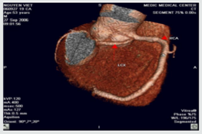
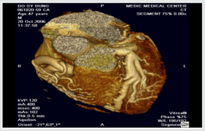
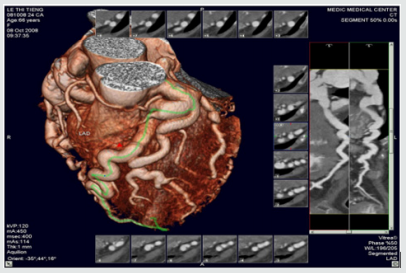
Lupine Publishers | MDCT in Diagnosis of Anomalies of Coronary Artery Origin and Course a Coronary MDCT-Angiographic study of 9572 patients

   Lupine Publishers | Advancements in Cardiology Research & Reports


Abstract

Background: Coronary anomalies are the causes of sudden cardiac deaths in young peoples, but usually asymptomatic. We perform this retrospective study to determine the types and prevalence of Coronary Anomalies of origin and course.

Method: The data of 9572 patients with Coronary CT-angiography by MDCT 640 Aquilion Toshiba machine were analyzed.

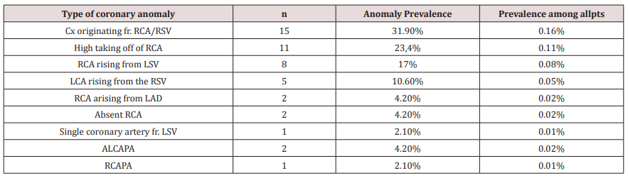
Results: Anomalous origin and course of coronary artery were detected in 47 (0.49%) of 9572 patients. The anomalous origins of Circumflex Artery from the RCA or the right sinus of Valsalva are most frequently visualized ( 15 pts [31.9%] ). High taking off of RCA observed in 11 pts ( 23.4% ).The RCA rising from the left sinus of Valsalva were seen in 8 pts ( 17% ).The Left Coronary Artery originates from the right sinus of Valsalva in 5 pts ( 10,6% ).The RCA arising from the LAD in 2pts (4,2% ).Absent RCA in 2 case (4.2%) and single coronary artery from LSV in one case (2.1%). The LCA rising from the Pulmonary Artery ( ALCAPA) in 2 cases and The RCA originating from the PA in one case ( RCAPA ).

Conclusion: Anomalies of coronary artery origin and course are rare but the diagnosis is very important to prevent SCD in young patients. MDCT with the Volume Rendered Images is the non-invasive modality that provides the valuable information to detect these anomalies.

Keywords: Multidetector Computed Tomography; Anomalies of coronary origin and course; sinus of Valsalva

Coronary artery anomalies are a diverse group of congenital heart diseases with manifestations and pathological mechanisms are highly variable. Coronary anomalies include anomalies of origin and course, anomalies of intrinsic coronary arterial anatomy like myocardial bridge, anatomy of coronary termination as coronary artery fistula and anomalous anastomotic vessels. Anomalies of coronary origin and course may associated with arrhythmias, myocardial infarction and sudden cardiac deaths in young people, especially on effort like athletes. We study 9572 patients with coronary MDCT-angiography to evaluate the type and the incidence of coronary anomalies of origin and course[1,2].

All patients who underwent coronary CT-angiography by MDCT 64O Aquilion Toshiba equipment ( IV contrast medium, gantry rotation of 0.33 msec, slice thickness 0.5mm ) in MEDIC HCMC Viet Nam, from January 2016 to January 2019 were included. The main indications of CT-angiography were acute coronary syndrome, stable angina, coronary CT-angiography prior to surgery, congenital heart diseases involving coronary artery...

The CT-angiograms with coronary anomalies were selected and analyzed. The anomalies of coronary origin and course were assessed [3-5].

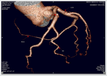
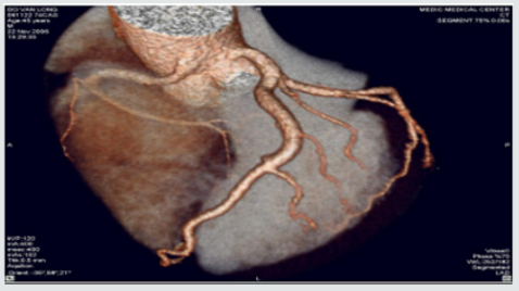
We included 9572 pts with anomalies of coronary origin and course based on results of CT-angiograms that were interpreted by two cardiologists. Anomalous origin and course of coronary artery were detected in 47 ( 0,49 %) of 9572 patients. The mean age of these pts was 63± 8.4, M/F=1.8 . The anomalous origins of Circumflex Artery from the RCA or the right sinus of Valsalva are most frequently visualized ( 15 pts [31.9%] ).High taking off of RCA observed in 11 pts ( 23.4% ) The RCA rising from the left sinus of Valsalva were seen in 8 pts ( 17% ).The Left Coronary Artery originates from the right sinus of Valsalva in 5 pts ( 10.6% ), in this subgroup, a patient presented by myocardial infarction resulting cardiac arrest was notified, the surgical re-implantation of LCA was performed .The RCA arising from the LAD in 2pts (4,2% ). Absent RCA in 2 case (4.2%) and single coronary artery from LSV in one case ( 2.1% ) (Table1 ).The Left Coronary Artery arising from the Pulmonary Artery ( ALCAPA ) in 2 cases ( 4.2% ) and The RCA originating from the PA ( RCAPA ) in one case ( 2.1% ). sinus of Valsalva (Figures 1-10).

Table 1.


Lupinepublishers-openaccess-cardiology

RSV: Right sinus of Valsalva, LSV: Left sinus of Valsalva, ALCAPA: Anomalous Left Coronary Artery from The Pulmonary Artery, RCAPA: Anomalous Origin of the Right Coronary Artery off The Pulmonary Artery.

Figure 1: Single coronary artery rising from LSV.


Lupinepublishers-openaccess-cardiology

This patient is of 52 ages, presented by atypical chest pain, the single coronary artery originating from LSV. The other case report of Prashanth Panduranga revealed the single coronary artery arising from RSV with exertional angina

Figure 2: High taking off of RCA.


Lupinepublishers-openaccess-cardiology

Some time causes myocardial infarction due to excessive angulation between RCA and Aorta. We have in our study one young patient of 24 y.o that had been transferred to the hospital by cardiac arrest , related to this anomaly. Operative re-implanted had been indicated to save the patient

Figure 3: RCA originates from LSV with intra-arterial course resulting Angina


Lupinepublishers-openaccess-cardiology

Figure 4: Anomalous origin of LCA from RSV


Lupinepublishers-openaccess-cardiology

Figure 5: RCA rising from LSV and Intra-arterial course of RCA.


Lupinepublishers-openaccess-cardiology

Figure 6:LCx arising from the RVS and Retro Aortic Course of LCx.


Lupinepublishers-openaccess-cardiology

Figure 7: LCx arising from the RVS and Retro Aortic Course of LCx.


Lupinepublishers-openaccess-cardiology

Figure 8: Anomalous Left Coronary Artery from The Pulmonary Artery.


Lupinepublishers-openaccess-cardiology

Figure 9: Other case of ALCAPA.


Lupinepublishers-openaccess-cardiology

Figure 10: Anomalous Origin of the Right Coronary Artery off The Pulmonary Artery.


Lupinepublishers-openaccess-cardiology
In our study, coronary anomalies of origin and course were detected in 47 of 9572 patients ( 0,49% ) that is consistent with the incidence of 0.27% to 1.66% reported in other series. The most frequent anomaly of origin and course was the Cx Artery arising from RCA/RSV ( 31.9% of anomaly prevalence and 0.16% among all patients ), this incidence is lower than previous published studies. The anomalies of origin and course of RCA were found in 17% and 4.2% respectively from LSV and LAD. This incidence is lower in comparison with previous study. Sudden deaths, myocardial infarction, arrhythmias related to the coronary anomalies were reported previously [6,7]. But these anomalies often asymptomatic, so early detection of coronary anomalies of origin and course is highly important. The former studies mainly based on the result of coronary angiography that is invasive modality. This study demonstrates MDCT is the noninvasive modality that provides important information related to coronary anatomy. Currently MDCT and MRI become fundamental to detection and diagnosis of coronary anomalies. Contrast enhanced ECG-gated 640-row MDCT coronary angiography is an accurate diagnostic method that can precisely detect the coronary anomalies of origin and course


For more Lupine Publishers Social Bookmarking Click on Below link

https://issuu.com/lupinepublishers-oaj/docs/lupine_publishers_social_bookmarking_sites.docx

Thursday, November 5, 2020

Lupine Publishers | Msct In Diagnosis of Congenital Heart Diseases in Viet Nam

Lupine Publishers | Advancements in Cardiology Research & Reports


Background: Congenital heart diseases associated with more malformations, complex aortopulmonary collaterals and anomalous coronary artery. Echocardiography is the initial diagnostic method but this method can be limited in complex congenital heart diseases.

Purpose: To assess the role of MDCT in congenital heart diseases (CHD) diagnosis compare with operative result and interventional angiography.

Methods: 910 patients with congenital heart diseases of 31.000 patients underwent cardiac angiography with 64 and 320 section CT at Medic Medical Center since 09/09/2006 to 30/12/2015.

Results: There are 658 operated cases, most of operated cases demonstrated the exact diagnosis of MDCT in congenital heart diseases.

Conclusions: MDCT is the fast and non-invasive diagnostic method with the high accuracy, overcomes the limit of echocardiography in complex congenital heart diseases diagnosis and provides the panorama and useful information’s prior to the operation.

Keywords: Congenital heart diseases; Cardiac multi-detector computed tomography, Multi-detector computed tomography in congenital heart diseases; Congenital heart diseases computed tomography

Congenital heart diseases effect ~ 1% of all live births in the general population. Complex congenital heart diseases associated with more malformations, complex aortopulmonary collaterals and anomalous coronary artery. Over the past few decades, the diagnosis and treatment of congenital heart diseases have greatly improved [1-6]. Diagnostic tools: X-ray, ECG, echocardiography, MRI and MDCT. ECG and X-Ray suggest the diagnosis but are not specific. Echocardiography is the initial diagnostic method for patients with suspected CHD but this method can be limited in complex CHD. The great capabilities of MRI for anatomic and functional assessment of the heart but MRI is time-consuming and may require patient sedation. Now enable CT to be used as an accurate noninvasive clinical instrument that is fast replacing invasive cine-angiography in the evaluation of CHD [1,2,5].

I. Improves both spatial and temporal resolution.

II. Increases scanning speed.

III. Improves diagnostic image quality by reducing respiratory artifacts

To assess the role of MDCT in congenital heart diseases (CHD) diagnosis compare with operative result and interventional angiography.

Subject: 910 patients with congenital heart diseases of 31.000 patients underwent cardiac angiography with 64 and 640 section CT at Medic Medical Center since 09/09/2006 to 30/12/2015.

Means and scanning techniques

a) Medic Medical Center scanned cardiac CT by 64 MDCT Toshiba Aquilion machine and Toshiba Aquilion One (320 MDCT), 0.5mm slice thickness, 0.5mm imaging reconstruction.

b) Two phases scanning: Don’t inject phase and contrast media injection phase: +Phase doesn’t inject contrast which help locate and assess coronary artery calcification.

c) +Phase inject contrast media: Medicine chasing phase and water chasing phase.

d) Contrast pumping machine is double-barreled Stellant (Medrad).

e) To inject contrast by intravenous right hand.

f) Contrast dose used 1mL/ kg.

g) Drug pump speed depends on patient status and disease.

h) Vitrea software: Reconstructed images by MPR, MIP and VRT.

i) Effective radiation dose is low (320-MDCT is 3.69±061mSv; 64-MDCT is 12-14mSv) (Figures 1).

Figure 1.


Lupinepublishers-openaccess-cardiology

a. The prospective study and case series report compare with operative and interventional angiography.

b. Data collection at the HCM city Heart Institute, Tam Duc Heart Hospital and Medic medical center (Figures 2-17).

Figure 2: Atrial septal defects and Ventricular septal defects.


Lupinepublishers-openaccess-cardiology

Figure 3: Patent ductus arteriosus.


Lupinepublishers-openaccess-cardiology

Figure 4: Coarctation of aorta.


Lupinepublishers-openaccess-cardiology

Figure 5: Double aortic arch.


Lupinepublishers-openaccess-cardiology

Figure 6: Tetralogy of Fallot.


Lupinepublishers-openaccess-cardiology

Figure 7: Pulmonary atresia with ventricular septal defect.


Lupinepublishers-openaccess-cardiology

Figure 8: Transposition of great vessels.


Lupinepublishers-openaccess-cardiology

Figure 9: Double outlet right ventricle.


Lupinepublishers-openaccess-cardiology

Figure 10: Single ventricle.


Lupinepublishers-openaccess-cardiology

Figure 11: Aortopulmonary window:


Lupinepublishers-openaccess-cardiology

Figure 12: Truncus arteriosus.


Lupinepublishers-openaccess-cardiology

Figure 13: Anomalous systemic venous return.


Lupinepublishers-openaccess-cardiology

Figure 14: Anomalous pulmonary venous connection.


Lupinepublishers-openaccess-cardiology

Figure 15: Single pulmonary artery.


Lupinepublishers-openaccess-cardiology

Figure 16: Pulmonary artery trunk aneurysm:


Lupinepublishers-openaccess-cardiology

Figure 17: Congenital pulmonary arteriovenous malformation.


Lupinepublishers-openaccess-cardiology

There are 658 operated cases, most of operated cases demonstrated the exact diagnosis of MDCT in congenital heart diseases.

Congenital heart diseases associated with more malformations, complex aortopulmonary collaterals and anomalous coronary artery. Echocardiography is the initial evaluative method for preand post-operation congenital heart diseases but this method can be limited in complex cases. Multi-detector computed tomography overcomes the limit of Echocardiography by multiplanar reconstruction (MPR) and volume rendered techniques (VRT) reconstruction . Volume rendered techniques (VRT) reconstruction clearly demonstrates the relationship between the heart and great vessels.

Multi-detector computed tomography is the fast and noninvasive diagnostic method with the high accuracy. Overcomes the limit of Echocardiography in complex congenital heart diseases. Provides the panorama and useful information’s prior to the surgery.

For more Lupine Publishers Social Bookmarking Click on Below link

https://issuu.com/lupinepublishers-oaj/docs/lupine_publishers_social_bookmarking_sites.docx

Interruption of the Aortic Arch in the Adult and Fulminant Myocarditis: A Strange Presentation

Introduction   53 years old female patient, who presented oppressive precordial pain, radiating to the neck and jaw, for which she went to...