Showing posts with label Journal of Cardiology. Show all posts

Saturday, June 19, 2021

Lupine Publishrers | Concomitant LVAD Implantation and Thoracic Surgery. Clinical Decision Making and Surgical Challenges

        Lupine Publishers | Advancements in Cardiology Research & Reports


Abstract

In the last years, left ventricular assist device (LVAD) indications have significantly broadened including candidates with multiple comorbidities even requiring combined surgical strategy. We present a case of a 56-year old patient affected by postischemic dilated cardiomyopathy in whom a lung nodule diagnosed during preoperative CT-scan could have contraindicated LVAD destination therapy. The patient underwent through a median sternotomy concomitant LVAD implantation on cardiopulmonary bypass followed by an atypical resection of the anterior part of the right lower lobe lung. A multidisciplinary, step-by-step approach to reduce the risk of right ventricular failure, bleeding and infections is presented.

Keywords: LVAD implantation; non-cardiac surgery; atypical lung resection

Indications to continuous flow left ventricular assist devices (CF-LVAD) have significantly broadened over the last decade considering even old patients with cardiac and extra-cardiac comorbidities [1]. Concomitant cardiac procedures to LVAD implantation are well described since nowadays up to 35% of implantations require concomitant cardiac surgery including valvular surgery, coronary artery bypass grafting, ventricular arrhythmias ablation, and atrial septal defects repair [1]. But the impact of these concomitant procedures is not well studied and guidelines are lacking. In particular for non-cardiac surgery experiences are limited to case series and the decision-making is driven on a single patient basis. To our knowledge, concomitant LVAD implantation and lung surgery has not been described before. We report the case of a patient who underwent a second generation CF-LVAD implantation followed by an atypical right lower lobe resection.

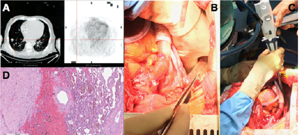
A 56-year-old man mildly obese and previous heavy smoker, presented with post-ischemic dilated cardiomyopathy and severely decompensated heart failure (orthopnea, dyspnea, fluid retention, and weight gain) with left ventricular ejection fraction (EF) of 17%. He also showed non-reversible post-capillary pulmonary hypertension and right ventricular (RV) dysfunction: Tricuspid Annular Plane Systolic Excursion (TAPSE) of 10 mm, RV Fractional Area Change (RVFAC) of 20%, Right Ventricular Stroke Work Indexed (RVSWI) of 400 mmHg/ml/m2 and a central venous pressure to wedge pressure ratio of 0.8. Few days after admission the patient developed hemoptysis. A 15-mm enhanced contrast lung nodule in the latero-basal segment of the right lower lobe (RLL) with a maximum standardized uptake value of 8, was demonstrated at the Positive Emission Tomography Scan (Figure 1A). The CT-scan showed a nodule suspicious for neuroendocrine tumor because of early contrast enhancement and a regular profile. Due to its peripheral position the nodule could not be reached with a transbronchial biopsy and a percutaneous computer tomography guided biopsy was considered to be too high risk. The pulmonary function tests were normal despite the presence of centrilobular and paraseptal emphysema.

Figure 1: (A)Computer Tomography and Positron Emission Tomography scan showing RLL enhanced contrast nodule; (B) manual palpation of lung nodule with deflated lung; (C) wedge resection of the RLL; (D) histology of the lung lesion showing a complex artero-venous malformation.

Lupinepublishers-openaccess-cardiology

He showed a positive clinical response to an aggressive diuretic therapy with significant reduction in weight and fluid retention, and improvement in right ventricular function (TAPSE 17 mm, RVFAC 33%, RVSWI 600 mmHg/mL/m2, central venous pressure to wedge pressure ratio of 0.4) which allowed to list the patient for an LVAD implantation. Considering right ventricular improvement, favorable position of the lung nodule and the complex coagulation management of a staged approach a combined procedure was preferred. A 24 hours infusion of levosimendan was completed in the immediate preoperative period. In consideration of the presence of the pulmonary lesion, we decided for a destination therapy configuration of the Jarvik 2000 (Jarvik Heart, Inc, New York, NY, USA) with an intraoperative retroauricolar implantation of the pedestal. After fixation of the pedestal, the pericardial space was approached via a median longitudinal sternotomy. Once the driveline was tunneled, LVAD implantation inside the left ventricle apex was performed on cardiopulmonary bypass (CPB) and beating heart, the outflow conduit was sutured to the ascending aorta. The progression from CPB to LVAD level 2 support was facilitated by continuous infusion of epinephrine (0.04 mcg/kg/min), norepinephrine (0.05 mcg/kg/min) and 20 ppm of inhaled nitric oxide to facilitate RV function.

When surgical and medical hemostasis were carefully achieved, one lung ventilation with 5 mL/Kg tidal volumes was initiated and a positive end expiratory pressure of 8 cmH2O, inhaled nitric oxide was increased to 30 ppm. Even if the PaO2/FiO2 ratio was 100 mmHg and the PaCO2 was 47 mmHg, the TEE showed normal RV function with a pulmonary artery systolic pressure of 40 mmHg. RLL exposure was achieved through the mediastinal pleura via the median sternotomic access. Manual palpation with inflated and noninflated lung allowed nodule identification and wedge resection of the RLL with an Echelon Flex 60 Endopatch (Ethicon Endo Surgery Inc., Cincinnati, OH) (Figure 1 B,C). Pathological examination demonstrated a complex artero-venous malformation (Figure 1D). The patient had an uneventful postoperative course with weaning from mechanical ventilation and inotropic support within the first 36 postoperative hours. He was discharged from the postoperative intensive care unit on day 6 and on day 17 to a rehabilitation center. Since the lung lesion was benign the patient was eligible to enter the heart transplantation list discharged and strictly monitored with a telemedicine program previously described [2].

The population of patients eligible for LVAD implantation has broadened over the last years and thanks to the reported improvements in mid-term outcomes and quality of life, the number of patients scheduled for a concomitant non-cardiac surgery intervention has progressively increased [3]. Planning a combined procedure in these fragile patients requires a thorough multidisciplinary approach, a strict preoperative medical optimization and the design of an effective and safe surgical procedure. Few cases of thoracic surgery in patients with CFLVAD have been published [4,5]. In this case, surgical plan was carefully defined in consideration of the major perioperative risks. Great attention was paid to RV function and preservation since hemodynamic changes following LVAD implantation are difficult to predict and have a huge impact on results [6,7]. Since no data have been published on the relationship between the amount of lung parenchyma excised and the variation of pulmonary resistances and thus of right ventricular afterload, we have chosen a step-bystep approach. An atypical lung resection was first performed. A RLL lobectomy would be a second surgical step only in case of demonstrated malignancy, minimizing in this way right ventricular impact and allowing a gradual adaptation of the RV to increased pulmonary resistances. During the procedure RV function was evaluated through surgical visual inspection, TEE and right heart catheterization allowing for immediate pump speed variations or pharmacological support titration.

The delicate interaction between RV, mechanical ventilation and hypoxia has been largely described in the population of patients suffering from acute respiratory distress syndrome and also during the intraoperative management of one lung ventilation for thoracic surgery, but it has never been studied in the context of LVAD support [8]. Indeed, RV disfunction after LVAD implantation is a life threating complication that can occur in 20 to 50% of patients [9] especially in the presence of high pulmonary vascular resistances. The RV showed a positive response to the increased venous return, to the geometrical adaptation of the interventricular septum and the augmented pulmonary vascular resistances. The favorable anatomical position of the lung nodule allowed to employ the median sternotomy for both LVAD implantation and lung resection. Concomitant procedure reduced the perioperative bleeding and infective risks. This case underlines the crucial role of a multidisciplinary approach, a preoperative medical optimization, a step-by-step surgical plan and a multimodal right ventricular evaluation.

For more Lupine Publishers Click on Below link

https://publons.com/publisher/7295/lupine-publishers-llc


Saturday, June 12, 2021

Lupine Publishers | Systematic Review of Gender Based Studies of Diagnosis and Treatment of Cardiovascular Disease in Last 20 Years

Lupine Publishers | Advancements in Cardiology Research & Reports


Abstract

Cardiovascular disease (CVD) encompasses a broad range of conditions. Coronary Artery Disease (CAD), commonly referred to as Ischemic Heart Disease (IHD), is the leading cause of death, morbidity, and mortality in the United State and women continue to have poorer outcomes than men. The causes of these discrepancies have yet to be fully elucidated. In this review, we reported gender-based studies of diagnosis, treatment, and outcome of CVD in the last 20 years.

Keywords: Cardiovascular disease; gender-based discrepancies; diagnosis; treatment

Introduction

Cardiovascular disease (CVD) is the major cause of death in the United State and other countries [1]. Age, ethnic, racial, and gender differences within CVD diagnosis and treatment have been well reported in several studies. Because men and women have different characteristics and predictive factors, decision-making based on the currently standardized frameworks and recommendations for clinical diagnosis and treatment tend to lead to poorer outcomes in women compared to men. In this paper, we present a short review of a few related works in gender-based CVD-related studies over the past 20 years.

Systematic Review in Last 20 years

In 1999, Kameneva MV et al. [2] reported that CVD mortality in men and menstrual women, particularly myocardial infarction (MI), is significantly higher than pre-menopausal women, which may be associated to the difference in age of red blood cells and the consequent differences in the structural features of menstrual and postmenopausal women. According to their results, men have hematocrite, blood viscosity, RBC aggregation, and RBC rigidity higher than pre-menopausal women. Jeanine E Roeters van Lennep et al. [3], concluded that diabetes, high density lipoproteins, and triglyceride levels were found to have a greater effect on women’s risk of coronary heart disease than men. They also found out that women and men show different responses to risk factors or to treatment in case of optimal treatment and prevention. In a research done by Edgar Argulian et al. [4], they suggested that women were more likely to experience coronary artery injury and bleeding problems than men, despite similar high angiographic and procedural success rates of PCI for both sexes. According to Regithz Zagrosek et al. [5], the increased prevalence of MetS in women over 40 years was 76% compared to 5 percent in men at the same age. In the cardiovascular risk associated with diabetes mellitus and hyperglycemia, the same gender difference was also observed.

In a study conducted by Kwok Leung Ong et al. [6], they suggested that sex hormones could be responsible for gender differences in coordination with blood pressure. Even though there was higher diastolic blood pressure in men, higher systolic pressure was reported in women which is a greater risk factor for CVD. They found that women had more prevalence of cardiovascular risk factors such as central obesity, high total cholesterol, and low HDL than men. Giusepp Mercuro et al. [7] reported significantly higher mortality rates among women with diabetes compared to men of the same age and women were less likely to ask for medical help. According to their report, women of older age have the nonischemic disease in patients with heart failure and have more severe symptoms of heart failure. By increasing the risk of a first acute myocardial infarction (AMI), smoking is more dangerous in younger women (< 50 years) than in men according to Maas AH et al. [8]. They also presented that Smoking also induces a downregulation of estrogen-dependent vasodilatation of the endothelial wall in young premenopausal women that can contribute to more problems in CHD patients. G Penno et al. [9] found that while CVD was more common in men, women had a greater risk profile for CVD and worse outcomes for therapy. They concluded that women were less likely to receive pharmacological therapy for hypertension hyperglycemia and dyslipidemia than men. In an exclusive analysis of a national cohort of CVD, Salim S. Virani et al. [10] found women were less likely to take statins and high-intensity statins than men did. Also, significant facility-level variation in both statin and highintensity statins used in female patients was observed.

Ramzi Y Khamis et al. [11] discovered that CVD diagnosis has been shown to be poorer since women with chest pain were less likely to perform ECG exercise and less probable to have coronary angiography. They stated that Females tend to experience CVD later in life and appear to be treated with less certainty than their male counterparts even when diagnosed. It has also been shown that women respond to antiplatelet agents differently from men, so residual platelet activity appears to be higher in them. According to Min Zhao et al. [12] report, women are worse at managing risk factors in CHD prevention. Results showed better regulation of blood pressure in women, but they had a poorer profile in glucose and lipid therapy target levels. Women tended to undergo less rigorous lipid-reduction therapy than men. Upon cardiovascular events, they were also less likely to take aspirin, ACE inhibitors, or statins. Niti R. Aggarwal et al. [13] suggested that considering advances in IHD mortality, it is still the leading cause of death among women. Compared to men, women are much less probable to be detected and involved in cardiac rehabilitation. Also, there are several non-traditional cardiac complications in women, including early menopause and menarche, gestational diabetes mellitus, and hypertension. In 2019, Amy Johnson et al. proved that women were more likely to die within one year of cardiac surgery and long-term follow-up than men. They also found that despite the fact that a higher mortality rate among women was correlated with remote MI, recent MI and previous PCI, there was a lower risk of death among men who had previous PCI [1].

Conclusion

The review of related works shows significant works and finding around risk factors and outcomes in CVD for men and women. But the causes of these discrepancies have yet to be fully addressed and require further research in this field of study to overcome poorer outcomes in women. Further detailed analysis is needed in order to design interventions and structures to minimize bias. The complication of pregnancy and other women-specific situations make it difficult to diagnose female patients with CVD in a timely manner.

For more Lupine Publishers Click on Below link

https://publons.com/publisher/7295/lupine-publishers-llc


Friday, February 12, 2021

Lupine Publishers | Epidemiological Aspects of Cardiac Decompensation Factors Renaissance Hospital N’Djamena Chad

Lupine Publishers | Advancements in Cardiology Research & Reports


Abstract


Introduction: Cardiac decompensation factors are numerous. Their identification allows better management of patients and limits the rate of rehospitalization. The aim of this work was to identify cardiac decompensation factors and improve their management at the renaissance hospital in N’Djamena, Chad.

Patients and Methods: This was a cross-sectional retrospective study conducted in the cardiology department at N’Djamena Renaissance Hospital, over a period of one year, from 01 January 2018 to 01 January 2019. All patients hospitalized for cardiac decompensation during this period and consented, were included.

Results: During the period of our study, 52 patients were included. The sex ratio was 1.9. The mean age was 48±9 years old. The predominant cardiovascular risk factors were arterial hypertension (37%, n = 19) and diabetes (27%, n = 14). The monthly income of our patients was in the majority of cases less than 200,000 FCFA (44%, n = 23). The main factors of cardiac decompensation were respectively, infections (18%, n = 9), supraventricular arrhythmias (16%, n = 8), changes in temperature (11%, n = 6), therapeutic nonobservance (11%, n = 6), dietary gap (10%, n = 5), and hypertensive relapses (10%, n = 5). The main etiologies of heart failure were ischemic cardiomyopathies (31%, n = 16), dilated cardiomyopathies (25%, n = 13), hypertensive cardiomyopathies (17%, n = 9), and rheumatic valvulopathies (15%). %, n = 8).

Conclusion: Rehospitalizations for cardiac decompensation are common after the first episode of hospitalization. The identification of the risk factors for this decompensation and their management make it possible to avoid these readmissions.

Keywords: Cardiac decompensation factors; Heart failure; Renaissance hospital N’Djamena characterized

Abbreviations: HF: Heart Failure; FFCA: Franc of the Financial Community in Africa; AFA: Atrial Fibrillation Arrhythmia; LV: Left Ventricle

Introduction

Heart failure (HF) is defined as a clinical syndrome characterized by chronic symptoms (dyspnea, fatigue) that may be accompanied by physical signs (crepitus, peripheral edema) caused by a structural cardiac abnormality and / or functional, resulting in decreased cardiac output [1]. It is a major public health problem because of its frequency and consequences in terms of morbidity and mortality and its economic impact on the health care system. Its prevalence is increasing because of the aging of the population but also because of the improvement of the management of many heart diseases including ischemic heart disease. It is one of the leading causes of hospitalization, morbidity and mortality, especially among the elderly [2]. Its evolution is clinically marked by periods of remission and exacerbation leading to recurrent hospitalizations. The number of readmissions for IC remains significant despite the therapeutic progress of recent years. The identification of decompensation factors and the optimization of their management could prevent these readmissions, particularly after hospitalization. The objective of this study was to identify cardiac decompensation factors and improve their management at the renal hospital in N’Djamena, Chad.

Patients and Methods

This was a cross-sectional retrospective study conducted in the cardiology department at N’Djamena Renaissance Hospital, over a period of one year, from 01 January 2018 to 01 January 2019. Were included all patients readmitted for cardiac decompensation during the study period and who gave their consent.

The parameters studied

Epidemiological characteristics: age, sex, cardiovascular risk factors (arterial hypertension, diabetes, chronic renal dysfunction with glomerular filtration rate <60ml/min / 1.73m2, obesity, dyslipidemia, alcohol, smoking), monthly cost of treatment in FFCA (1 US dollar = 593,720 FFCA). Clinical features: cardiac decompensatory factors (difference in diet, unsuitable exercise, temperature, alcohol, AFA, other rhythm disorders, hypertensive pressure, ischemic episode, anemia, bronchopulmonary infection, other infection, renal failure, poor compliance drug, hyperthyroidism, untreated sleep apnea syndrome), etiologies of HF. Electrocardiographic characteristics: arrhythmias (atrial or ventricular extrasystoles, atrial fibrillation or flutter, atrial tachycardia, ventricular tachycardia), repolarization abnormalities, sinoatrial or atrioventricular blocks. Echocardiographic features: dilated cardiac cavities, wall hypertrophy, diastolic dysfunction of the left ventricle (LV); abnormalities of left ventricular kinetics (hyperkinesia, hypokinesia, akinesia), LV systolic dysfunction (systolic ejection fraction <45%), valvular abnormality, congenital anomaly, pulmonary arterial hypertension.

Statistical Analysis

In this study, a descriptive statistical analysis was applied using Microsoft Excel, quantitative variables were presented by their mean and standard deviation and qualitative variables were by percentages.

Ethics

This work was done by obtaining the approval of the hospital management and the consent of the patients.

Results

During the period of our study, 52 patients were included. Men predominated with 65% of cases (n = 34). The sex ratio was 1.9. The average age was 48±9 years old with a minimum age of 22 years and a maximum age of 79 years. Patients were educated only in 38% of cases (n = 20). The predominant cardiovascular risk factors were hypertension (19%), diabetes (14%), dyslipidemia (11%), and 19%). The monthly income of our patients was in the majority of cases less than 200,000 FCFA. The rates were, respectively, between CFAF 100,000 and 200,000 in 44% of cases (n =23) and less than CFAF 100,000 in 33% of cases (n = 17). Only 12 patients (23%) had an income above 200,000 FCFA. The health insurance rate was observed in 13% of cases (n=7). (Table 1) shows the characteristics of the patients. The main factors of cardiac decompensation are shown in Table 2. The most frequent were respectively, infections in 18% of cases (n = 9), including 4 cases of bronchopulmonary infections (8%), supraventricular rhythm disorders in 16% of cases (n=8) including 5 cases of AFA (10%), changes in temperature especially heat with 6 cases (11%), poor therapeutic compliance 6 cases (11%), the difference diet in 10% of cases (n = 5), and hypertensive outbreaks in 10% of cases (n=5). The most common etiologies of IC (Table 3) were ischemic cardiomyopathies in 31% of cases (n=16), of which 4 patients (8%) had benefited from myocardial revascularization, dilated cardiomyopathies in 25% (n=16). = 13), hypertensive cardiomyopathies in 17% of cases (n =9) and rheumatic valvulopathies in 15% (n=8). Other etiologies were less frequent in this series such as post-embolic pulmonary heart in 6% of cases (n=3), congenital heart disease in 4% (n=2) and pericardial affections in 2% of cases (n= 1).

Table 1: Patient Characteristics.

Lupinepublishers-openaccess-cardiology

Table 2: Cardiac Decompensation Factors.

Lupinepublishers-openaccess-cardiology

Table 3: Etiologies of HF.

Lupinepublishers-openaccess-cardiology

Discussion

The post-hospitalization period is conducive to rehospitalization, but long-term chronic HF patient follow-up is important because the morbidity and mortality rate remains high in this group of patients, even though many treatments have been shown to be effective. The follow-up of these patients makes it possible to optimize the therapeutics, the monitoring and to detect early the signs of decompensation. Cardiac rehabilitation plays an important role in the management of HF and should be part of the modern strategy for the management of patients with stable heart failure. This includes not only physical training, but also rehabilitation of drug treatments, control of risk factors, psychological management and finally, patient education [3]. The search for this factor that shifts a situation of balance during a decompensation is fundamental because its treatment can be very profitable. Several standards for HF support highlight the need to look for a triggering factor [4]. We studied the importance of triggers for cardiac decompensation in a population of readmitted patients who were followed for chronic HF. The main triggering factors identified were, in order of frequency, infections, atrial fibrillation, therapeutic nonobservance, temperature variations, mainly heat, uncontrolled hypertension, diet deviation, anemia, myocardial ischemia. These factors are globally found in the literature data. In this study, the first triggering factor found was the existence of an infection (18%) with bronchopulmonary infections (8%) at their head. They are promoted by decreasing the effectiveness of coughing, bronchiolar elasticity, efficiency of the mucociliary system and swallowing disorders.

The mechanisms involved in cardiac decompensation are multiple. Several studies have highlighted the important role of infections, particularly bronchopulmonary infections, in cardiac decompensation, especially in elderly patients [5-8]. The second triggering factor was a supraventricular rhythm disorder (16%), especially ACFA (10%). Suppression of atrial systole results in increased LV filling pressures and promotes the onset of HF thrust. AFA is common in HF and its prevalence increases with the severity of CI [9-13]. Cardiac decompensation was attributed to noncompliance in 8% of the cases in this study, but adherence remains difficult to assess. This non-compliance was favored by several factors in our patients; the high number of drugs with their adverse effects, given the high rate of comorbidities observed (diabetes, high blood pressure, kidney failure), the economic level which was low in almost 50% of patients and the absence of health insurance, the level of education and the influence that might have on the understanding of the disease. The therapeutic nonobservance in the HF varies in the literature from 10 to 99% according to the evaluation method used [14]. Compliance is responsible for an increase in the number of hospitalizations and a worsening of clinical signs [15]. A multidisciplinary intervention has shown its effectiveness on adherence to 30-day treatment [16] and an educational intervention has improved compliance and decreased re-hospitalization rates [17]. Anemia was found in 4 patients in our series.

This result is weak compared to data from the literature because, according to several authors, anemia is frequent during IC and is a poor prognostic factor in chronic HF [18,19], increased clinical signs, aggravation of functional status [20]. However, there is little data on the involvement of acute anemia in cardiac decompensation. In addition, the management of the etiology of HF remains essential to prevent complications and readmissions and to improve the prognosis. In our study we found that in coronary patients (31%) only 8% had benefited from myocardial revascularization, valvular patients (15%) and those with congenital heart disease (4%) had not received reparations. surgical procedures that were necessary. In our context, the identification of the decompensation factors must be of paramount importance for the clinician and should allow the improvement of the management of the pathology in particular at the preventive level. Hence the importance of educating the patient and those around him. The establishment of appropriate structures and care networks for heart failure in all regions of Chad will allow better monitoring of patients with rapid access to the specialist physician.

The limits of our work

Our study presents several methodological limitations. First, it is a retrospective study with information gathered from the reports that sometimes did not contain all the necessary data. The size of our sample is small, we will have to lead other more representative studies in the future. And many patients have not received etiologic treatment of HF.

Conclusion

Rehospitalizations for cardiac decompensation are common after the first episode of hospitalization. The identification of the risk factors for this decompensation and their management make it possible to avoid these readmissions. Hence the importance of emphasizing access to care with appropriate therapeutic means, regular monitoring and therapeutic education.

For more Lupine Publishers Click on Below link

https://www.zoominfo.com/c/lupine-publishers-llc/43353514


Friday, October 30, 2020

Lupine Publishers | The Left Common Carotid Artery Rises from the Main Pulmonary Artery

  Lupine Publishers | Advancements in Cardiology Research & Reports


Abstract

A young female patient of 15y.o presented at my hospital by dyspnea on effort and palpitation for one year. Mental deficiency was notified. Physical examination detected a 3/6 continuous murmur at the 2ndRICS. In the past history, PDA had been suspected by her physician, associated with recurrent bronchitis. Trans-thoracic Echocardiography showed an enlarged LV of 57mm with normal EF of 69% , LCA=5mm, RCA=3.5mm at origin, no suspected sign of PDA was seen. Only a continuous flow was visualized in the PA. CT-Angiography with IV contrast medium showed the Left Common Carotid Artery rising from the Pulmonary Artery trunk. PDA was not presented. The Left Common Carotid Artery then was re-implanted into the aortic arch normally with a favorable postoperative progress.

Keywords: Carotid Artery; Pulmonary Artery; Anomalous origin

Anomalous origin of the left common carotid artery is very rare and has been reported previously. We present an operated case of this topic with clinical finding, cardiac ultrasound and MDCT imaging.

Figure 1: Right aortic Arch.


Lupinepublishers-openaccess-cardiology

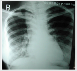
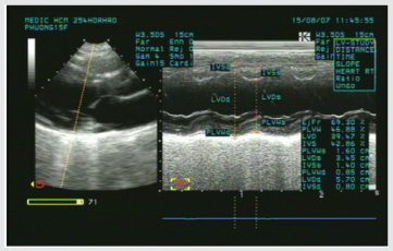
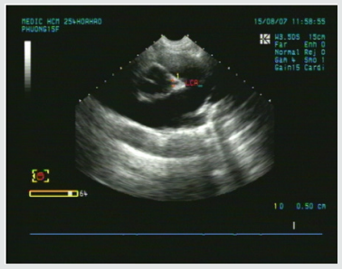
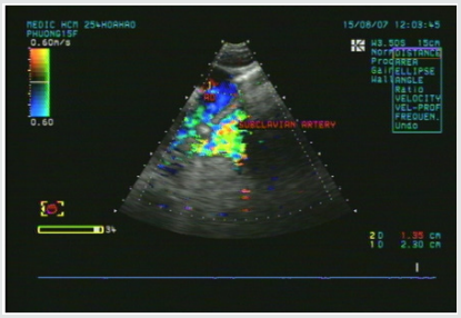
A young female patient of 15y.o presented at my hospital by dyspnea and palpitation when running and fast walking for one year. Mental deficiency was notified, she had some difficulties to learn at school. Physical examination detected a 3/6 continuous murmur at the 2ndRICS. In the past history, PDA has been suspected by her physician, associated with recurrent bronchitis. Her body state was normal with 1m60 of height and 48 kg of weight. She was evaluated immediately by a chest X ray that showed a right aortic arch (Figure 1). The trans-thoracic echocardiography that revealed an enlarged LV of 57mm with normal EF of 69% (Figure 2), LCA=5mm, RCA=3.5mm at origin (Figure 3). No suspected sign of PDA was detected except a continuous flow presented in the Pulmonary Artery (Figure 4).

Figure 2: Enlarged LV& normal systolic function.


Lupinepublishers-openaccess-cardiology

Figure 3: Normal LCA at origin.


Lupinepublishers-openaccess-cardiology

Figure 4: Continuous flow in the PA.


Lupinepublishers-openaccess-cardiology

Figure 5: Absence of aortic origin of the LCCA.


Lupinepublishers-openaccess-cardiology

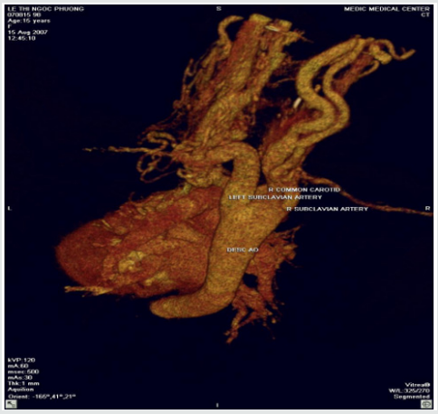
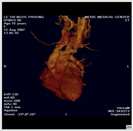
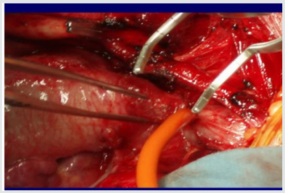
CT-Angiography (MDCT 64) with IV contrast medium Ultravist, slice thickness=1mm visualized a right aortic arch, aberrant origin of the left subclavian artery, dilatation of the branches rising from aortic arch with increased collateral vessels (Figure 5). Especially, MDCT 64 showed the Left Common Carotid Artery ( LCCA ) rose from the PA trunk (Figure 6) PDA was not detected. Patient underwent uncomplicated surgical repair: the Left Common Carotid Artery was re-implanted into the aortic arch normally with a favorable post-operative progress (Figure 7).

Figure 6: The LCCA rising from the Main PA roof.


Lupinepublishers-openaccess-cardiology

Figure 7: Re-implantation of the LCCA.


Lupinepublishers-openaccess-cardiology

Anomalous origin of the Left Common Carotid Artery from the Pulmonary Artery Trunk has been previously reported as rare cases. Kagami Mijaji et al. [1] has reported a case of anomalous origin of the Artery from the Right Pulmonary Artery. Onyekachukwu et al. [2] has described a case of anomalous origin of the Left Common Carotid Artery from the Main Pulmonary Artery. In this article, my patient was not infant with CHARGES syndrome that includes multiple congenital anomalies like the patients in their reports. She was a teenage patient without other congenital disease. The role of ultrasound is orienting for the indication of Computed Tomography or DSA. In case of present turbulent flow in the PA, Coronary Fistula and other shunts from the head and neck vessels should be considered [3].

Anomalous origin of the Left Common Carotid Artery is very rare congenital defect that maybe isolated or associated with some syndromes. Noninvasive diagnostic methods as Ultrasound and CTA may confirm the diagnosis and inform the anatomical relation of the anomalous vessels prior to operate


For more Lupine Publishers Social Bookmarking Click on Below link

https://issuu.com/lupinepublishers-oaj/docs/lupine_publishers_social_bookmarking_sites.docx

Monday, June 24, 2019

Lupine Publishers - Journal of Cardiology Research


Psychopathy of the careerist is a developmental disorder marked by emotional deficits and an increased risk for antisocial behavior. It is not equivalent to the diagnosis Antisocial Personality Disorder, which concentrates only on the increased risk for antisocial behavior and not a specific cause-ie, the reduced empathy and guilt that constitutes the emotional deficit. Our review considers data regarding the neurobiology of this disorder. Dysfunction within the amygdala’s role in reinforcement learning and the role of ventromedial frontal cortex in the representation of reinforcement value is stressed. Data is also presented indicating potential difficulties within parts of temporal and posterior cingulate cortex. Suggestions are made with respect to why these deficits lead to the development of the disorder. Knowledge of recent neuorobiology is proving our thesis that Darwin was wrong when formulated his theorem„ Survival of the fittest“. Reality in 21 century is showing that “Survival of the careerist“ based on the Quantum Entanglement Entropy (QEE) is more valid principle of Social Dynamics in our days. Careeristic Competition is the main cause of the QEE leading to icreased complications through Coincidenses of Social Dynamics - Lupine Publishers.

For more Lupine Publishers Open Access Journals Please visit our website


For more Journal of Cardiology Research articles Please Click Here:
https://www.lupinepublishers.com/cardiology-journal/

Wednesday, April 24, 2019

Journal of Cardiology & Clinical Research-Lupine Publishers


Ward Clinical pharmacy, pharmaceutical care, and consultant pharmacist service can be considered interesting instruments in the world of cognitive service to improve global clinical/economical results in multi-disciplinary medical team. This pharmacist field of working can be considered as an opportunity in this time in with a great number of new drugs molecules, medical - diagnostic procedure added to the more complexity of cure, polytherapy and more necessity to containment cost for drugs and medical devices than past. Reduction of medication therapy errors is needed and actively requested by patients and health organization institution and government. Multi-professional healthcare team is the right keywords in today health- care systems. Clinical pharmacist contributes today in many disciplines like oncology, toxicology, emergencies medicine, ICU, nuclear medicine, infectious disease, nephrology, nutrition service and other - Lupine Publishers.


For more Lupine Publishers Open Access Journals Please visit our website

Interruption of the Aortic Arch in the Adult and Fulminant Myocarditis: A Strange Presentation

Introduction   53 years old female patient, who presented oppressive precordial pain, radiating to the neck and jaw, for which she went to...