Lupine Publishers | Advancements in Cardiology Research & Reports
Abstract
Background: Congenital heart diseases associated with more malformations, complex aortopulmonary collaterals and
anomalous coronary artery.
Echocardiography is the initial diagnostic method but this method can be limited in complex congenital heart diseases.
Purpose: To assess the role of MDCT in congenital heart diseases (CHD) diagnosis compare with operative result and
interventional angiography.
Methods: 910 patients with congenital heart diseases of 31.000 patients underwent cardiac angiography with 64 and 320
section CT at Medic Medical Center since 09/09/2006 to 30/12/2015.
Results: There are 658 operated cases, most of operated cases demonstrated the exact diagnosis of MDCT in congenital heart
diseases.
Conclusions: MDCT is the fast and non-invasive diagnostic method with the high accuracy, overcomes the limit of
echocardiography in complex congenital heart diseases diagnosis and provides the panorama and useful information’s prior to the
operation.
Keywords: Congenital heart diseases; Cardiac multi-detector computed tomography, Multi-detector computed tomography in
congenital heart diseases; Congenital heart diseases computed tomography
Introduction
Congenital heart diseases effect ~ 1% of all live births in the
general population. Complex congenital heart diseases associated
with more malformations, complex aortopulmonary collaterals
and anomalous coronary artery. Over the past few decades, the
diagnosis and treatment of congenital heart diseases have greatly
improved [1-6]. Diagnostic tools: X-ray, ECG, echocardiography, MRI
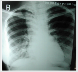
and MDCT. ECG and X-Ray suggest the diagnosis but are not specific.
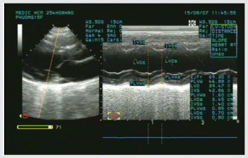
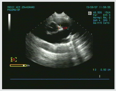
Echocardiography is the initial diagnostic method for patients with
suspected CHD but this method can be limited in complex CHD. The
great capabilities of MRI for anatomic and functional assessment
of the heart but MRI is time-consuming and may require patient
sedation. Now enable CT to be used as an accurate noninvasive
clinical instrument that is fast replacing invasive cine-angiography
in the evaluation of CHD [1,2,5].
I. Improves both spatial and temporal resolution.
II. Increases scanning speed.
III. Improves diagnostic image quality by reducing respiratory
artifacts
Purpose
To assess the role of MDCT in congenital heart diseases (CHD)
diagnosis compare with operative result and interventional
angiography.
Material and Methods
Subject: 910 patients with congenital heart diseases of 31.000
patients underwent cardiac angiography with 64 and 640 section
CT at Medic Medical Center since 09/09/2006 to 30/12/2015.
Means and scanning techniques
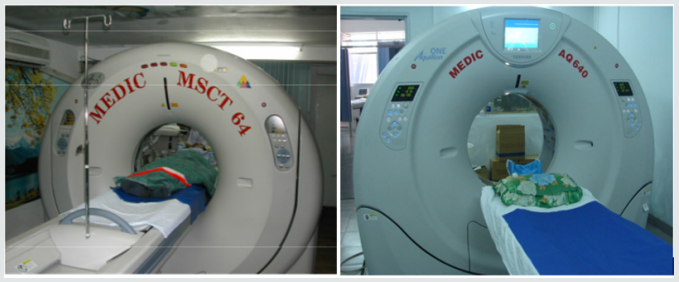
a) Medic Medical Center scanned cardiac CT by 64 MDCT
Toshiba Aquilion machine and Toshiba Aquilion One (320
MDCT), 0.5mm slice thickness, 0.5mm imaging reconstruction.
b) Two phases scanning: Don’t inject phase and contrast
media injection phase: +Phase doesn’t inject contrast which
help locate and assess coronary artery calcification.
c) +Phase inject contrast media: Medicine chasing phase and
water chasing phase.
d) Contrast pumping machine is double-barreled Stellant
(Medrad).
e) To inject contrast by intravenous right hand.
f) Contrast dose used 1mL/ kg.
g) Drug pump speed depends on patient status and disease.
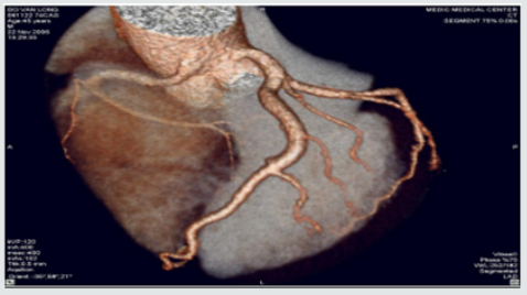
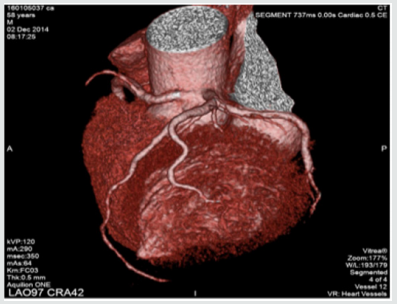
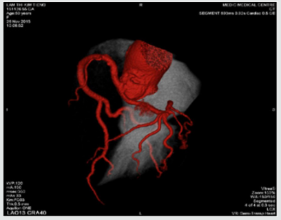
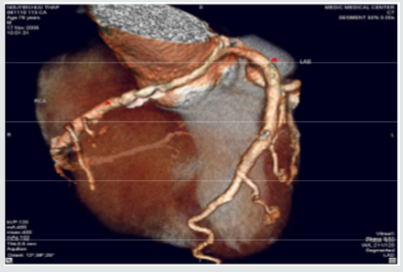
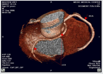
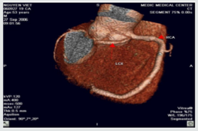
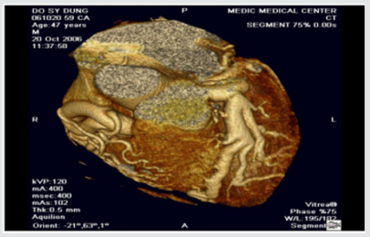
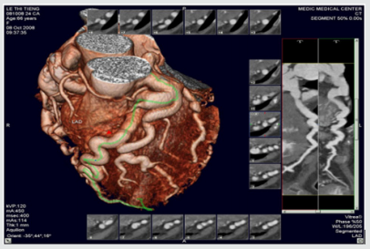
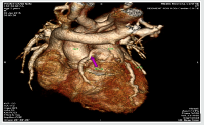
h) Vitrea software: Reconstructed images by MPR, MIP and
VRT.
i) Effective radiation dose is low (320-MDCT is
3.69±061mSv; 64-MDCT is 12-14mSv) (Figures 1).
Figure 1.
Data analysis
a. The prospective study and case series report compare
with operative and interventional angiography.
b. Data collection at the HCM city Heart Institute, Tam Duc
Heart Hospital and Medic medical center (Figures 2-17).
Figure 2: Atrial septal defects and Ventricular septal defects.
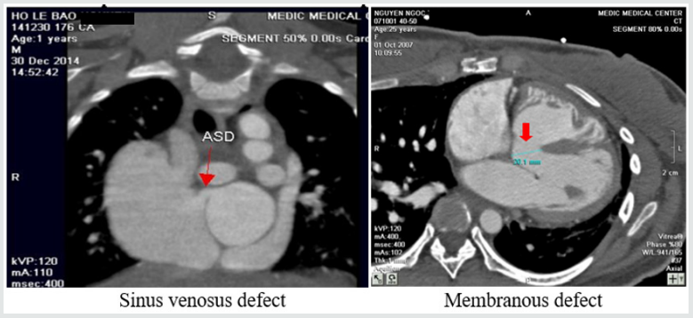
Figure 3: Patent ductus arteriosus.
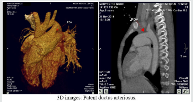
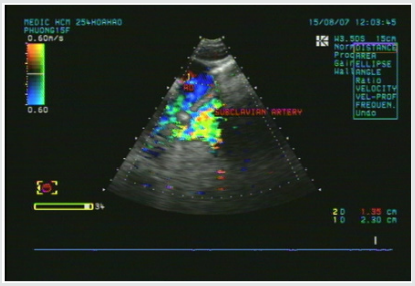
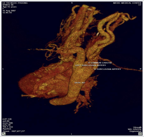
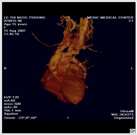
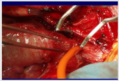
Figure 4: Coarctation of aorta.
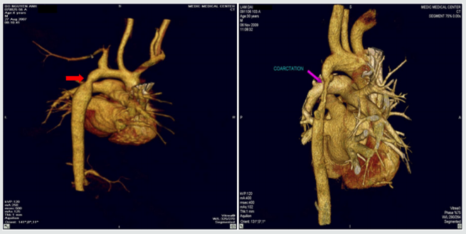
Figure 5: Double aortic arch.
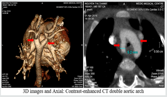
Figure 6: Tetralogy of Fallot.
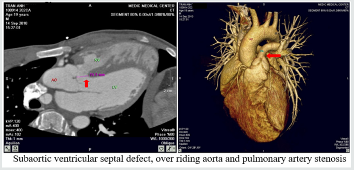
Figure 7: Pulmonary atresia with ventricular septal defect.
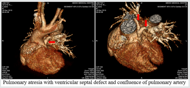
Figure 8: Transposition of great vessels.
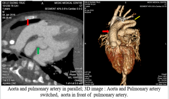
Figure 9: Double outlet right ventricle.
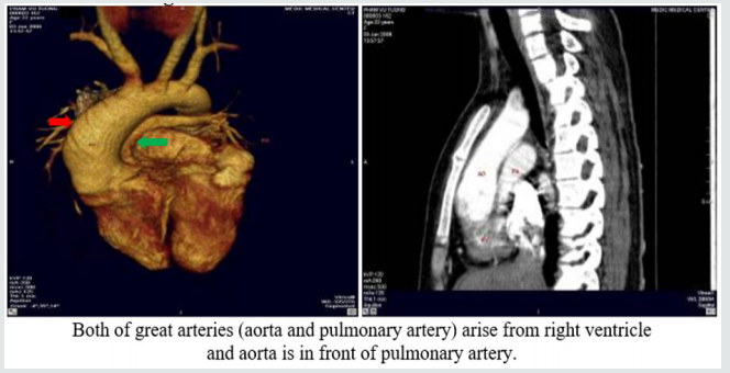
Figure 10: Single ventricle.
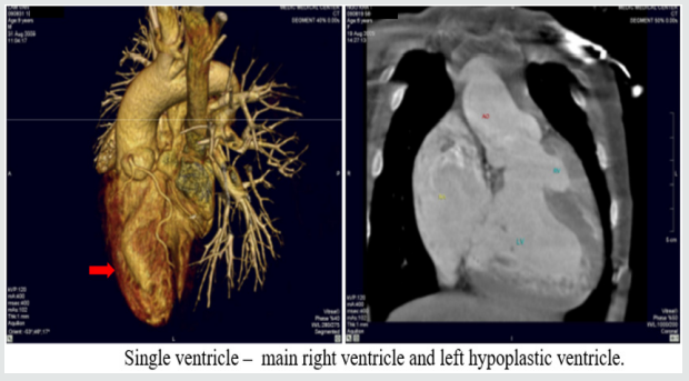
Figure 11: Aortopulmonary window:
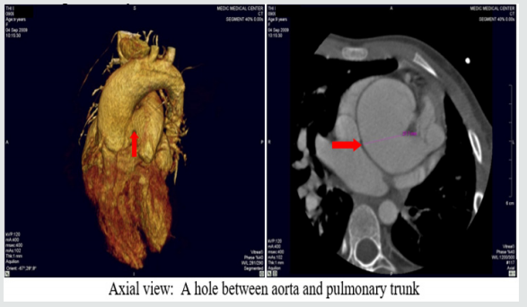
Figure 12: Truncus arteriosus.
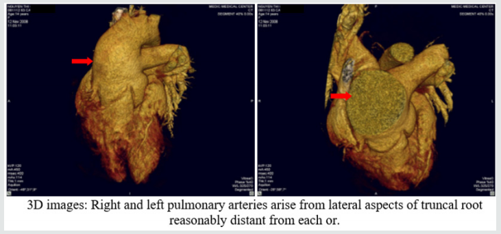
Figure 13: Anomalous systemic venous return.
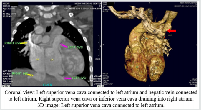
Figure 14: Anomalous pulmonary venous connection.
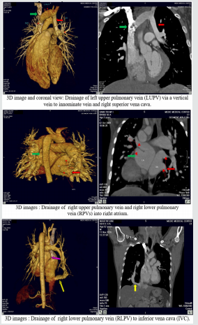
Figure 15: Single pulmonary artery.
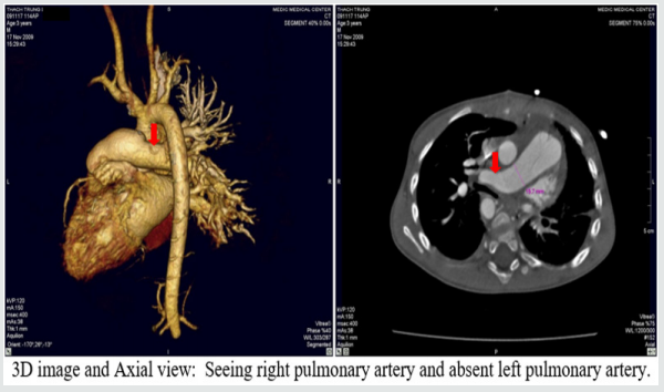
Figure 16: Pulmonary artery trunk aneurysm:
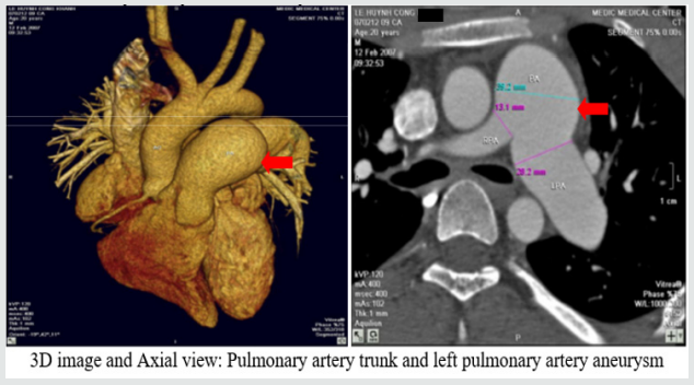
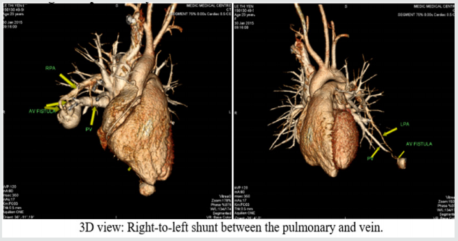
Figure 17: Congenital pulmonary arteriovenous malformation.
Results
There are 658 operated cases, most of operated cases
demonstrated the exact diagnosis of MDCT in congenital heart
diseases.
Discussion
Congenital heart diseases associated with more malformations,
complex aortopulmonary collaterals and anomalous coronary
artery. Echocardiography is the initial evaluative method for preand
post-operation congenital heart diseases but this method can
be limited in complex cases. Multi-detector computed tomography
overcomes the limit of Echocardiography by multiplanar
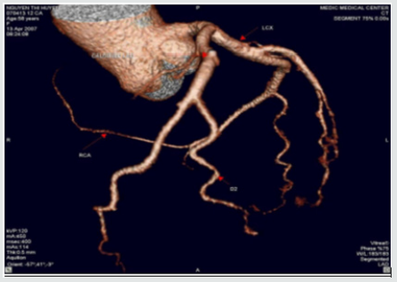
reconstruction (MPR) and volume rendered techniques (VRT)
reconstruction . Volume rendered techniques (VRT) reconstruction
clearly demonstrates the relationship between the heart and great
vessels.
Conclusion
Multi-detector computed tomography is the fast and noninvasive diagnostic method with the high accuracy. Overcomes the
limit of Echocardiography in complex congenital heart diseases.
Provides the panorama and useful information’s prior to the
surgery.
For more Lupine Publishers Social Bookmarking Click on Below link
https://issuu.com/lupinepublishers-oaj/docs/lupine_publishers_social_bookmarking_sites.doc