Friday, January 22, 2021

Lupine Publishers | Relationship between Depression and Physical Activity of Myocardial Infarction Patients after Treatment

Lupine Publishers | Advancements in Cardiology Research & Reports


Abstract


Physical activity is a secondary prevention that can reduce mortality and re-admission in patients with myocardial infarction. The objective of this study was to identify the relationship with physical activity of myocardial infarction patients after treatment. This study used a cross sectional method. A total of 150 myocardial infarction patients were selected using a purposive sampling technique. The results showed that the majority of STEMI post-treatment patients have mild physical activity (82%). There is also a significant relationship between depression and the level of physical activity of myocardial infarction patients after treatment (p = 0.003), OR = 0.144 (95% CI; 0.032-0.635). Depression in myocardial infarction patients at the time of the attack, if not intervened properly, it will persist and affect physical activity after treatment. A recommendation is directed to the nursing department to assess depression in patients with newly diagnosed of myocardial infarction.

Keywords: Physical activity; myocardial infarction; depression in myocardial infarction patients

Background

Physical activity is recommended by the European Society of Cardiology (ESC) as a long-term therapy in prevention for myocardial infarction patients Ibanez et al. [1]; Amsterdam et al. [2]. Physical activity can reduce mortality, re-admission and improve the quality of life of patients with myocardial infarction Andersen & Laustsen, [3]; Dalal, et al. [4]; Ek et al. [5]. Although physical activity is recommended as a long-term therapy in STEMI patients, only 37% of patients actively engage in physical activity after treatment Mckee et al. [6]. Several factors are known to have an association with physical activity in myocardial infarction patients, one of which is depression Mckee et al. [6]. Depression in myocardial infarction patients occurs 48-72 hours after a heart attack Kala, et al. [7]. Depression has a negative effect on post-treatment recovery in myocardial infarction patients, and causes lower compliance to treatment programs Homma et al. [8]; Kumar et al. [9]. The objective of this study was to identify the relationship between depression and physical activity of patients with myocardial infarction after treatment.

Method

This design of the study was a cross sectional study. Sampling was carried out using non-probability sampling techniques with a sample of 150 people. The inclusion criteria in this study were patients aged ≥18 years who were diagnosed with myocardial infarction. The study was conducted at the regional hospital of Jambi province, Indonesia in February - March 2018. Data collection was done using PHQ-9 Patient Depression Questionairre Kroenke et al. [10] and International Physical Activity Questionnaire (IPAQ) Strath et al. [11].

Findings

Characteristics of Respondents

Of the 150 post-treatment STEMI patients, the majority of patients were aged 18-60 years (73.3%), were male (78.7%), and 75.3% of whom had passed 7 to 30 days post-treatment. The majority of respondents were in the category of mild depression 69.3%, whereas the rest experienced moderate-severe depression, and 82% of respondents were at the level of physical activity with mild categories. Relationship between depression and physical activity of post-treatment myocardial infarction patients. The results of the analysis of the relationship between depression and physical activity showed that 95.7% of post-treatment myocardial infarction patients experienced moderate-severe depression with mild physical activity. Meanwhile, among post-treatment myocardial infarction patients who experience mild depression, 24% had moderate-heavy physical activity. Fisher exact test results obtained p = 0.003, so it can be concluded that there is a relationship between depression and physical activity. From the results of the analysis also demonstrated that the value of OR is 0.144 (95% CI; 0.032-0.635). By looking at the OR values it can be concluded that post-treatment myocardial infarction patients who experience mild depression would have a 0.144 times greater chance of having moderate-heavy physical activity compared to patients who have moderate-severe depression category

Discussion

Physical activity is a key component in heart disease patients that is beneficial in reducing the risk of relapse Thompson et al. [12]. In this study, the results of the analysis showed that 82% of respondents are at the level of mild physical activity. The results of this study are similar to studies conducted by Matthias, 2017 in Sri Lanka, where 56, 7% of respondents had low physical activity Matthias et al. [9]. Low physical activity is a trigger for the occurrence of myocardial infarction. Physical activity increases the process of arteriosclerosis formation, decreases inflammation, and triggers the formation of thrombosis Cheng et al. [13]. Many factors can affect physical activity. The study of Mckee et al. [6]. concluded that depression was one of the dominant factors causing low physical activity. This is the same as the results of this study. The results of bivariate testing found a relationship between depression and physical activity. Patients with myocardial infarction who experience depression tend to smoke, have low physical activity, and consume a lot of alcohol Qing Wu et al. [14]. In addition, experience during an attack is a cause of depression, and this continues for up to two months after the attack. This state of depression results in the patient becoming silent and limiting their physical activity.

Conclusion

Post-treatment myocardial infarction patients have a mild level of physical activity, and depression during the attack still occurs in myocardial infarction patients after undergoing treatment in the hospital. Depression, if not properly intervened, will cause changes in physical activity of myocardial infarction patients after treatment.

For more Lupine Publishers Click on Below link

https://www.zoominfo.com/c/lupine-publishers-llc/433535148

Monday, January 11, 2021

Lupine Publishers | MDCT in Diagnosis of Anomalies of Coronary Artery Origin and Course a Coronary MDCT-Angiographic study of 9572 patients

Lupine Publishers | Advancements in Cardiology Research & Reports


Abstract

Background: Coronary anomalies are the causes of sudden cardiac deaths in young peoples, but usually asymptomatic. We perform this retrospective study to determine the types and prevalence of Coronary Anomalies of origin and course.

Method: The data of 9572 patients with Coronary CT-angiography by MDCT 640 Aquilion Toshiba machine were analyzed.

Results: Anomalous origin and course of coronary artery were detected in 47 (0.49%) of 9572 patients. The anomalous origins of Circumflex Artery from the RCA or the right sinus of Valsalva are most frequently visualized ( 15 pts [31.9%] ). High taking off of RCA observed in 11 pts ( 23.4% ).The RCA rising from the left sinus of Valsalva were seen in 8 pts ( 17% ).The Left Coronary Artery originates from the right sinus of Valsalva in 5 pts ( 10,6% ).The RCA arising from the LAD in 2pts (4,2% ).Absent RCA in 2 case (4.2%) and single coronary artery from LSV in one case (2.1%). The LCA rising from the Pulmonary Artery ( ALCAPA) in 2 cases and The RCA originating from the PA in one case ( RCAPA ).

Conclusion: Anomalies of coronary artery origin and course are rare but the diagnosis is very important to prevent SCD in young patients. MDCT with the Volume Rendered Images is the non-invasive modality that provides the valuable information to detect these anomalies.

Keywords: Multidetector Computed Tomography; Anomalies of coronary origin and course; sinus of Valsalva

Introduction

Coronary artery anomalies are a diverse group of congenital heart diseases with manifestations and pathological mechanisms are highly variable. Coronary anomalies include anomalies of origin and course, anomalies of intrinsic coronary arterial anatomy like myocardial bridge, anatomy of coronary termination as coronary artery fistula and anomalous anastomotic vessels. Anomalies of coronary origin and course may associated with arrhythmias, myocardial infarction and sudden cardiac deaths in young people, especially on effort like athletes. We study 9572 patients with coronary MDCT-angiography to evaluate the type and the incidence of coronary anomalies of origin and course[1,2].

Methods

All patients who underwent coronary CT-angiography by MDCT 64O Aquilion Toshiba equipment ( IV contrast medium, gantry rotation of 0.33 msec, slice thickness 0.5mm ) in MEDIC HCMC Viet Nam, from January 2016 to January 2019 were included. The main indications of CT-angiography were acute coronary syndrome, stable angina, coronary CT-angiography prior to surgery, congenital heart diseases involving coronary artery...

The CT-angiograms with coronary anomalies were selected and analyzed. The anomalies of coronary origin and course were assessed [3-5].

Results

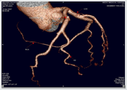
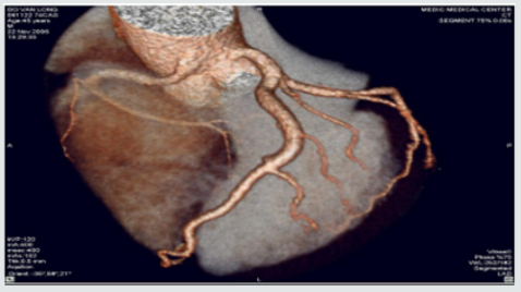
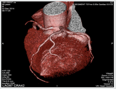
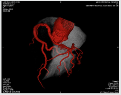
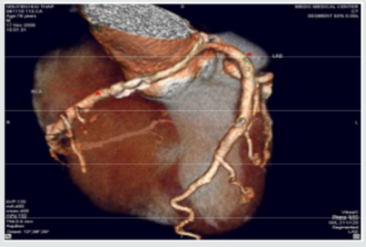
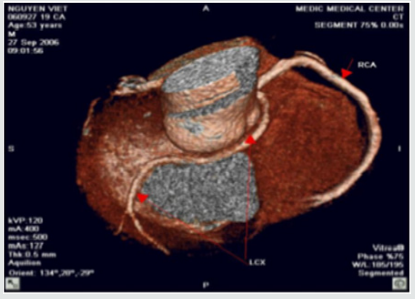
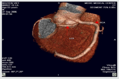
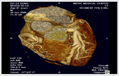
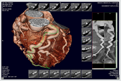
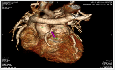
We included 9572 pts with anomalies of coronary origin and course based on results of CT-angiograms that were interpreted by two cardiologists. Anomalous origin and course of coronary artery were detected in 47 ( 0,49 %) of 9572 patients. The mean age of these pts was 63± 8.4, M/F=1.8 . The anomalous origins of Circumflex Artery from the RCA or the right sinus of Valsalva are most frequently visualized ( 15 pts [31.9%] ).High taking off of RCA observed in 11 pts ( 23.4% ) The RCA rising from the left sinus of Valsalva were seen in 8 pts ( 17% ).The Left Coronary Artery originates from the right sinus of Valsalva in 5 pts ( 10.6% ), in this subgroup, a patient presented by myocardial infarction resulting cardiac arrest was notified, the surgical re-implantation of LCA was performed .The RCA arising from the LAD in 2pts (4,2% ). Absent RCA in 2 case (4.2%) and single coronary artery from LSV in one case ( 2.1% ) (Table1 ).The Left Coronary Artery arising from the Pulmonary Artery ( ALCAPA ) in 2 cases ( 4.2% ) and The RCA originating from the PA ( RCAPA ) in one case ( 2.1% ). sinus of Valsalva (Figures 1-10).

Table 1.

Lupinepublishers-openaccess-cardiology

RSV: Right sinus of Valsalva, LSV: Left sinus of Valsalva, ALCAPA: Anomalous Left Coronary Artery from The Pulmonary Artery, RCAPA: Anomalous Origin of the Right Coronary Artery off The Pulmonary Artery.

Figure 1: Single coronary artery rising from LSV.

Lupinepublishers-openaccess-cardiology

This patient is of 52 ages, presented by atypical chest pain, the single coronary artery originating from LSV. The other case report of Prashanth Panduranga revealed the single coronary artery arising from RSV with exertional angina

Figure 2: High taking off of RCA.

Lupinepublishers-openaccess-cardiology

Some time causes myocardial infarction due to excessive angulation between RCA and Aorta. We have in our study one young patient of 24 y.o that had been transferred to the hospital by cardiac arrest , related to this anomaly. Operative re-implanted had been indicated to save the patient

Figure 3: RCA originates from LSV with intra-arterial course resulting Angina

Lupinepublishers-openaccess-cardiology

Figure 4: Anomalous origin of LCA from RSV

Lupinepublishers-openaccess-cardiology

Figure 5: RCA rising from LSV and Intra-arterial course of RCA.

Lupinepublishers-openaccess-cardiology

Figure 6:LCx arising from the RVS and Retro Aortic Course of LCx.

Lupinepublishers-openaccess-cardiology

Figure 7: LCx arising from the RVS and Retro Aortic Course of LCx.

Lupinepublishers-openaccess-cardiology

Figure 8: Anomalous Left Coronary Artery from The Pulmonary Artery.

Lupinepublishers-openaccess-cardiology

Figure 9: Other case of ALCAPA.

Lupinepublishers-openaccess-cardiology

Figure 10: Anomalous Origin of the Right Coronary Artery off The Pulmonary Artery.

Lupinepublishers-openaccess-cardiology

Discussion & Conclusion

In our study, coronary anomalies of origin and course were detected in 47 of 9572 patients ( 0,49% ) that is consistent with the incidence of 0.27% to 1.66% reported in other series. The most frequent anomaly of origin and course was the Cx Artery arising from RCA/RSV ( 31.9% of anomaly prevalence and 0.16% among all patients ), this incidence is lower than previous published studies. The anomalies of origin and course of RCA were found in 17% and 4.2% respectively from LSV and LAD. This incidence is lower in comparison with previous study. Sudden deaths, myocardial infarction, arrhythmias related to the coronary anomalies were reported previously [6,7]. But these anomalies often asymptomatic, so early detection of coronary anomalies of origin and course is highly important. The former studies mainly based on the result of coronary angiography that is invasive modality. This study demonstrates MDCT is the noninvasive modality that provides important information related to coronary anatomy. Currently MDCT and MRI become fundamental to detection and diagnosis of coronary anomalies. Contrast enhanced ECG-gated 640-row MDCT coronary angiography is an accurate diagnostic method that can precisely detect the coronary anomalies of origin and course.


For more Lupine Publishers Click on Below link

https://www.zoominfo.com/c/lupine-publishers-llc/433535148


Interruption of the Aortic Arch in the Adult and Fulminant Myocarditis: A Strange Presentation

Introduction   53 years old female patient, who presented oppressive precordial pain, radiating to the neck and jaw, for which she went to...